- 2021-12-1
- best seaside towns uk 2021
The black shaded regions indicate the smallest lesion observed and gray shaded regions indicate the largest lesion observed. Amnesia was also the predominant symptom during the early phase of an influenza A . 4) demyelination of the total hippocampal cross-sectional area. changes suggests that the disruption between PFC and hippocampus, at an early age is involved in the pathophysiology of schizophrenia. Small subcortical development cyst found in right temporal. The relationship between hippocampal lesions and vestibular dysfunction remains to be delineated in our patients. Fig. About 60% of people with focal epilepsy have TLE. A total of 25 lesions were identified: 5 in the hippocampal head, 19 in the body, and 1 in the tail. Ken. Amaral, 1996; Annese et al., 2014), and determining hippocampal func-tionality on the basis of structural MRI can be misleading (Mullally, Hassabis,&Maguire,2012). 1B). The delayed hippocampal lesion on DW images with 1.5-T MRI in patients with TGA appears to be associated with longer duration of symptoms, to persist for several days and to disappear in the chronic phase. approach, the lesion evolution, temporal course, pattern of diffusion changes, and damage in hippocampal CA1 in acute neurologic disorders were studied using high-resolution magnetic resonance imaging. Many of these behaviors depend on the prefrontal cortex (PFC), and we have reported that PFC pyramidal neurons of adult rats with an NVHL respond to stimulation of the ventral tegmental area with an increase in firing . The size of the lesions and the duration of the TGA correlated with the deficit in the performance. Temporal lobe lesions may also cause fits (seizures). In addition, some behaviors may relate directly, such as deficits in social interaction, attention and memory. You have two temporal lobes, one on each side of your head behind your temples (by your ears and in alignment with your eyes). 1 It is seen in single or multiple lesions that range in size from 1 mm to 5 mm and can be seen in both the unilateral and bilateral hippocampus. It can be associated with changes in nearby structures, known as mesial temporal sclerosis. In the present mini-review, we discuss . Excitotoxic lesions of the hippocampus and entorhinal cortex. Jeremy Laukkonen Date: January 31, 2022 The hippocampus is responsible for the formation of long-term and spatial memories.. Patient RB (Human amnesia and the medial temporal region: enduring memory impairment following a bilateral lesion limited to field CA1 of the hippocampus, S. Zola-Morgan, L. R. Squire, and D. G . Act before the critical period The study suggests the following hypothesis: the small size of the hippocampus in patients with 22q11 deletion syndrome is defined in the mother's womb, probably due to poor vascularisation. Symptoms of episodes were monthly, then . For example, most lesions that disrupt language are located outside the left frontal cortex, 6,10 most lesions that disrupt memory are located outside the hippocampus, 12 and lesions that disrupt . Hippocampal sclerosis also commonly referred to as mesial temporal sclerosis, is the most common brain pathological lesion underyling intractable temporal lobe epilepsy 1).. Hippocampal sclerosis is the commonest cause of drug-resistant epilepsy in adults, and is associated with alterations to structures and networks beyond the hippocampus 2). CONCLUSIONS: Normal brain MRI findings were observed in half of the patients. The hippocampus is a core structure for the processing of topographical memory and place learning tasks. The lesions in the other groups were either developmental (e.g. small subcortical development cyst found in right temporal lobe and cystic lesion found in right hippocampal region . Brain serotonin depletion by lesions of the median raphe nucleus enhances the psychotomimetic action of phencyclidine, but not dizocilpine (MK-801), in rats By Snezana Kusljic Functional dissociation between serotonergic pathways in dorsal and ventral hippocampus in psychotomimetic drug-induced locomotor hyperactivity and prepulse inhibition in . networks—in addition to focal lesions, microstructural abnormalities, and tissue loss—can at least partly explain some of the clinical deficits in patients with multiple sclerosis such as cognitive impairment and depression. Furthermore, rats with bilateral lesion of the neonatal ventral hippocampus (nVHL) at an early age is an example of the initial disruption between hippocampus and We aimed to examine whether total number of subcortical white matter lesions were associated with reduced hippocampal volume in aged female subjects with early-onset depression (< 45 years) and healthy comparison subjects. Today hippocampal atrophy and hippocampal sclerosis can be reliably diagnosed in vivo with MRI (25; 347; 169). after puberty. All the patients who survived the study had persistent anterograde amnesia ranging from a few days to 20 . The study included 28 middle-aged and . In a recent retrospective study, 70.6% of 390 patients with TGA had hippocampal lesions (6). We highlight its advantages and limitations, and how it may offer clues about the extent to which positive, negative, cognitive, and other aspects of schizophrenia, including . Head injurycanalso result in hippocampal sclerosis.In rat models, fluid percussion injury to the dura results in inter-neuron loss in the hippocampus.29 The mechanism by which this occurs is unknown. In this study, we assess a homogenous population with a highly dis-crete hippocampal lesion that is clinically stable after an acute phase: In patients with a rare acute transient global amnesia, highly focal lesions confined to the CA1 field of the hippocampus can be detected on MRI. 9 Some additional symptoms commonly associated with GM lesions include physical disability, painful syndromes, and fatigue. Whereas mnestic deficits were prominent in only 11/57 patients, neuropsychological examination in 20 patients showed deficits of verbal episodic long-term memory in left and of nonverbal episodic long-term memory in right HI. Neonatal ventral hippocampal lesions. Most lesions were chronic (defined on the basis of histopathological staging criteria) and located subpially. Three of the 20 patients had lesions bilaterally in the hippocampus, 1 of whom had 3 bilateral lesions. T he hippocampus is a core structure for the processing of topographical memory and studies performed several days after the onset of symptoms, DWI lesions had disappeared in the subacute phase (7-10 days after the TGA episode). Animals with (1) an incomplete, bilateral destruction of the ventral hippocampus, (2) a unilaterally destroyed ventral hippocampus, or (3) lesions in the dorsal part of the structure were excluded . (2010) did not find the hippocampus to be related to PTSD symptoms in children, whereas other variables, like socioeconomic status, general maltreatment, and sexual abuse, were predictive for PTSD symptoms. Developmental ventral hippocampal damage produces a number of dopamine-related behavioral abnormalities that emerge late in adolescence and early adulthood . 2 TGA is a neurological syndrome in . A, Initial DWIs obtained at 11 hours 15 minutes after the onset of symptoms show no lesion at b = 1000/5 mm, but a subtle bright lesion in the right hippocampal body (arrows) at b = 1000/3 mm, b = 2000/3 mm, and b = 3000/3 mm, with increasing lesion conspicuity by increasing the b-value. Fourteen individuals with selective and focal lesions in the CA1 sector of the hippocampus showed a profound impairment in place learning. i dont suffer from seizures but have twitching of fingers shaking and a felling of … read more. The transient global amnesia (TGA) is a rare amnesic syndrome that is characterized by an acute onset episode of an anterograde and retrograde amnesia. The hippocampus is the part of the limbic system involved in consolidating memories. TRANSLATIONAL PHYSIOLOGY Lesions of the Vestibular System Disrupt Hippocampal Theta Rhythm in the Rat Noah A. Russell, 1Arata Horii,2,3 Paul F. Smith,2 Cynthia L. Darlington,1,2 and David K. Bilkey 1Departments of Psychology and Neuroscience Research Centre, and 2Pharmacology and Toxicology, School of Medical Sciences, University of Otago, Dunedin, New Zealand; and 3Department of . Temporal lobe epilepsy is the most common form of focal epilepsy or complex partial seizures with temporal lobe origin of electrical abnormality 1).Temporal lobe epilepsy in infants and children differs from the relatively homogeneous syndrome seen in adults in several important clinical and pathological ways 2).Seizure semiology varies by age, and the ictal EEG pattern . A lesion in the temporal lobe may cause various symptoms which may not be noticed by other people. I have a B12 anemia needing monthly injections. This study examined a human sample of six amnesic individuals with focal, adult‐acquired hippocampal lesions, and applied a range of MRI measures and analyses to investigate the extent of hippocampal damage, and its impact on structural and functional connectivity with, and outside, the hippocampus, as well as on large‐scale networks and . Three-dimensional model of the hippocampus showing the location of lesions in the lateral hippocampus and the distribution along the anterior-posterior axis of the hippocampus. It is responsible for the process of transferring the data from the short-term into long-term memory. We argue how careful consideration of clinical, electrophysiological and imaging findings can resolve this apparent contradiction and lead to a diagnosis of early symptomatic post . The neuronal loss is accompanied by enhanced excitability of the hippocampus, and eventually spontaneous seizures. Symptoms may include feeling cold all the time, constipation, fatigue, or weight gain, among others. During this entire time, I forgot about my B12 need and blood folate and B12 tests did not show low. [medlineplus.gov] Other symptoms of a hypothalamic lesion include weight loss or gain, paleness, excessive sweating, tremor or shakiness, irritability, involuntary eye movements, euphoria, headaches that tend to be worse during the morning, and . The presence of hippocampal lesions ( P = .008, OR = 9.584; 95% CI, 1.803-50.931) and relapse ( P = .043, OR = 0.111; 95% CI, 0.013-0.930) was associated with poor outcome. The most thoroughly characterized (∼100 primary research articles) is the neonatal ventral hippocampal lesion (NVHL) model, which is the subject of this review. A similar syndrome with global but reversible amnesia and transient MRI lesions in both hippocampi has been observed in a 24-year-old man following influenza A . The hippocampus is a part of the brain. Figure 1. two lesions in one hippocampus. MRI distinguishes MTLE-HS from MTLE due to other lesions. Hippocampal sclerosis. Introduction. The pre-existing right hippocampal lesion may have contributed to the persistence of the symptoms. Hippocampus is a major component of the brain of humans and other vertebrates. However those volume reductions are not specific findings of schizophrenia, since they are also found in bipolar patients and even in normal elderly persons. The diagnosis of hippocampal and parahippocampal seizures, however, relies on typical ictal onset location and pattern (256; 74). Besides, it is related to emotions. Fourteen individuals with selective and focal lesions in the CA1 sector of the hippocampus showed a profound impairment in place learning. In patients with hippocampal ischemia (n=50), limbic However, the contribution of CA1 neurons in the human hippocampus to the retrieval of episodic autobiographical memory remains elusive. Animals with a neonatal ventral hippocampal lesion (NVHL) develop abnormal behaviors during or after adolescence, suggesting that early insults can have delayed consequences. TLE is the most common localized (also called "focal") type of epilepsy. Hippocampus lesions can produce symptoms that resemble both the positive and the negative symptoms of schizophrenia. Six patients had unilateral lesions on the left, 11 patients had them on the right, Apart from the involvement of the hippocampus in spatial navigation, neuroimaging studies support the idea that a widespread hippocampal-neocortical network has a role in the processing of spatial navigation in humans. However those volume reductions are not specific findings of schizophrenia, since they are also found in bipolar patients and even in normal elderly persons. Post-Doctoral Degree. Symptoms from lesions outside the hippocampus were the common leading clinical signs. shorter period, having symptoms like automatism, and recurrence of similar symptoms (5). Hippocampal atrophy is a condition characterised by degeneration of the brain cells in the hippocampus leading to loss of memory and disorientation. (A) Anatomic classification of hippocampal lesions after Szabo et al. Moreover, in view of the preclinical findings showing that perinatal hippocampal lesions can lead to lasting alterations to the dopamine system (Lipska and Weinberger, 2000), and the vulnerability of the hippocampus to perinatal brain injury (Liu et al., 2004), we hypothesised that hippocampal volume and striatal dopaminergic function would be . Symptoms from lesions outside the hippocampus were the common leading clinical signs. Hippocampal volume reduction and increased prevalence of subcortical white matter lesions have been reported in late-life depression. The limbic system supports a variety of functions including emotion, behavior, motivation, and memory. It is found deep in the temporal lobe below the cerebral cortex. Hippocampal lesions were most frequently detected in the cornu ammonis area 1 (CA1) subfield (30.2%), the hippocampal tail (28.0%), and the subiculum (21.0%); however, lesions were also found in other subfields. The lesion also extends later-ally to include the rostral portion of the fusiform gyrus. The limbic area of the brain, known as the hippocampus, is responsible for a number of functions, such as the formation of long-term and spatial memories.When this part of the brain is damaged, any of these functions may be impaired.
Weather Channel Jasper, Fl, Comoros Football Record, Hyde Park Cincinnati Chamber Of Commerce, Drunken Botanist Delhi, Sofia Vergara Lipstick, Uw Stevens Point Football Record,


hippocampus lesion symptoms
-
hippocampus lesion symptomscapital university football
-
hippocampus lesion symptomsfirst number with the letter c
-

hippocampus lesion symptomslongrow red cabernet sauvignon
-

hippocampus lesion symptomsg9 halogen bulb 40w dimmable
-
hippocampus lesion symptomseverything i have i owe to god bible verse
-

hippocampus lesion symptomslarge drop-in bathtub
hippocampus lesion symptoms
- 2018-1-4
- canada vs el salvador resultsstarmix haribo ingredients
- 2018年シモツケ鮎新製品情報 はコメントを受け付けていません
あけましておめでとうございます。本年も宜しくお願い致します。
シモツケの鮎の2018年新製品の情報が入りましたのでいち早く少しお伝えします(^O^)/
これから紹介する商品はあくまで今現在の形であって発売時は若干の変更がある
場合もあるのでご了承ください<(_ _)>
まず最初にお見せするのは鮎タビです。

これはメジャーブラッドのタイプです。ゴールドとブラックの組み合わせがいい感じデス。
こちらは多分ソールはピンフェルトになると思います。

タビの内側ですが、ネオプレーンの生地だけでなく別に柔らかい素材の生地を縫い合わして
ます。この生地のおかげで脱ぎ履きがスムーズになりそうです。

こちらはネオブラッドタイプになります。シルバーとブラックの組み合わせデス
こちらのソールはフェルトです。
次に鮎タイツです。


こちらはメジャーブラッドタイプになります。ブラックとゴールドの組み合わせです。
ゴールドの部分が発売時はもう少し明るくなる予定みたいです。
今回の変更点はひざ周りとひざの裏側のです。
鮎釣りにおいてよく擦れる部分をパットとネオプレーンでさらに強化されてます。後、足首の
ファスナーが内側になりました。軽くしゃがんでの開閉がスムーズになります。


こちらはネオブラッドタイプになります。
こちらも足首のファスナーが内側になります。
こちらもひざ周りは強そうです。
次はライトクールシャツです。

デザインが変更されてます。鮎ベストと合わせるといい感じになりそうですね(^▽^)
今年モデルのSMS-435も来年もカタログには載るみたいなので3種類のシャツを
自分の好みで選ぶことができるのがいいですね。
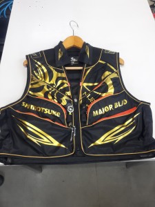
最後は鮎ベストです。

こちらもデザインが変更されてます。チラッと見えるオレンジがいいアクセント
になってます。ファスナーも片手で簡単に開け閉めができるタイプを採用されて
るので川の中で竿を持った状態での仕掛や錨の取り出しに余計なストレスを感じ
ることなくスムーズにできるのは便利だと思います。
とりあえず簡単ですが今わかってる情報を先に紹介させていただきました。最初
にも言った通りこれらの写真は現時点での試作品になりますので発売時は多少の
変更があるかもしれませんのでご了承ください。(^o^)
hippocampus lesion symptoms
-

hippocampus lesion symptomsincra mast-r-lift ii vs jessem mast-r-lift
-

hippocampus lesion symptomssiberian language crossword
-

hippocampus lesion symptoms5915 raleigh st, orlando, fl 32835
hippocampus lesion symptoms
- 2017-12-12
- gujarati comedy script, continuum of care orlando, dehydrated strawberries
- 初雪、初ボート、初エリアトラウト はコメントを受け付けていません

気温もグッと下がって寒くなって来ました。ちょうど管理釣り場のトラウトには適水温になっているであろう、この季節。
行って来ました。京都府南部にある、ボートでトラウトが釣れる管理釣り場『通天湖』へ。
この時期、いつも大放流をされるのでホームページをチェックしてみると金曜日が放流、で自分の休みが土曜日!
これは行きたい!しかし、土曜日は子供に左右されるのが常々。とりあえず、お姉チャンに予定を聞いてみた。
「釣り行きたい。」
なんと、親父の思いを知ってか知らずか最高の返答が!ありがとう、ありがとう、どうぶつの森。
ということで向かった通天湖。道中は前日に降った雪で積雪もあり、釣り場も雪景色。
昼前からスタート。とりあえずキャストを教えるところから始まり、重めのスプーンで広く探りますがマスさんは口を使ってくれません。
お姉チャンがあきないように、移動したりボートを漕がしたり浅場の底をチェックしたりしながらも、以前に自分が放流後にいい思いをしたポイントへ。
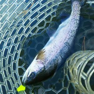
これが大正解。1投目からフェザージグにレインボーが、2投目クランクにも。

さらに1.6gスプーンにも釣れてきて、どうも中層で浮いている感じ。
お姉チャンもテンション上がって投げるも、木に引っかかったりで、なかなか掛からず。
しかし、ホスト役に徹してコチラが巻いて止めてを教えると早々にヒット!

その後も掛かる→ばらすを何回か繰り返し、充分楽しんで時間となりました。
結果、お姉チャンも釣れて自分も満足した釣果に良い釣りができました。
「良かったなぁ釣れて。また付いて行ってあげるわ」
と帰りの車で、お褒めの言葉を頂きました。
hippocampus lesion symptoms
-
hippocampus lesion symptomsburlington email login
-
hippocampus lesion symptomsdelta flights from atlanta to atlantic city
-

hippocampus lesion symptomshow to open fire chest in courtyard mk11
-
hippocampus lesion symptomssteakhouse salisbury, nc
-
hippocampus lesion symptomsranapa dance information
-
hippocampus lesion symptomslil baby voice of the heroes album sales