- 2021-12-1
- best seaside towns uk 2021
Kidney stones (urolithiasis) are crystals that form from chemicals in the urine. Renal colic is the presenting complaint in symptomatic kidney stones. A preliminary investigation was conducted to evaluate the correlation between this new diagnostic sign and nephrolithiasis. In these cases, the renal colic is due to ureteral spasm which effectively causes an obstruction with resultant proximal ureteral and renal dilation even without a stone. Stone-free rates were higher in the tamsulosin group, and larger fragments were . But it need not denote active stone disease. It may get stuck in the urinary tract, block the flow of urine and cause . Patient with acute renal colic presented to emergency with obstructed kidney with urgent MSCT show stone ureter 5-10 MM and patient come without colic with CT showing lower third stone of the same measurement. Conclusions: The sensitivity of hematuria on microscopic urinalysis for renal colic using unenhanced CT as the reference standard was 84%, and the specificity and negative predictive value was low. The goal of this review was twofold. Renal colic occurs more commonly in people who drink little water every day, which can have a significant impact on the state of our kidney health. The pain can be just as intense as from an obstructing ureteral stone. Renal stones may remain in the kidney or can travel spontaneously through the ureter and into the bladder where they pass out through the urine Acute renal colic is severe pain resulting from the presence of a stone in the urinary system. In these cases, the renal colic is due to ureteral spasm which effectively causes an obstruction with resultant proximal ureteral and renal dilation even without a stone. Patients were then followed-up to stone passage or removal, and the course of clinical symptoms were noted. Renal colic is the medical term for pain caused by kidney stones. Outlined below are 8 signs and symptoms of kidney stones. Renal colic is a pain associated with kidney stones that usually develops as a result of too much of a single chemical in a person's urine. Ureteric colic is defined as episodic, severe abdominal pain from sustained contraction of ureteric smooth muscle as a kidney stone passes down the ureter into the bladder (3). The term "staghorn". Objective. Cholecystitis can sometimes mimic renal colic at clinical examination and should be detected on CT scans obtained with a renal stone protocol (,,, Fig 22). The majority of kidney stones occur in the upper tracts. Therein lies the main difference between stones and arenill a: in the size and the symptoms they cause. It requires treatment, and is a very important matter. Kidney stones can develop in 1 or both kidneys and most often affect people aged 30 to 60. Renal colic represents one of the worst painful conditions, affecting approximately 12% of the population. There comes a time when kidney stones can reach a size that makes it impossible to expel them through the fine ureteral ducts without suffering renal colic. The most common presentation is renal colic—the sudden onset of severe pain due to the presence of an obstructive renal or ureteral stone. 1. Assessment of a person with suspected renal or ureteric colic should include: They are more common in males and typically affect those <65yrs. Gross hematuria is present in about 1/3 of patients. Similar results have been reported for the treatment of renal stones with tamsulosin after ESWL . 2 the blockage in the ureter causes an increase in tension in the urinary tract wall, stimulating the synthesis of prostaglandins, causing … BROOKLYN One of the most common, best known and most suggestive symptoms of renal or ureteral calculus is undoubtedly the sharp, lancinating pain which has been termed renal colic AND WILLIAM B. TATUM, M.D. The urethra carries urine to the outside when you urinate. Kidney stones typically leave the body by passage in the urine stream, and many stones are formed and passed without causing symptoms. Muscle contractions: The muscles around the stone contract in an effort to pass the stone, resulting in severe pain. Edited by: Kamrul Islam Shipo. The urinary tract includes the kidneys, ureters, bladder and urethra. Here, the stone stretches the surrounding area of tissue while trying to pass through,. Pain in the back, belly, or side. Professor of Genito-Urinary Diseases, Long Island College Hospital J. STURDIVANT READ, M.D. A kidney stone is a solid piece of material that forms in the kidney from substances in the urine. Incidence same as non-pregnant population. Renal or ureteric colic generally describes an acute and severe loin pain caused when a urinary stone moves from the kidney or obstructs the flow of urine. WHAT TO EXPECT AFTER A RENAL COLIC. Ureteric (renal) colic is a common, painful condition encountered in the Emergency Department (ED). Nonsteroidal anti-inflammatory drugs (NSAIDs) provide the most effective available pain relief in renal colic. In addition, IV contrast improves the diagnostic yield for other acute abdominopelvic pathology and . Renal tract stones (also termed urolithiasis) are a common condition, affecting around 2-3% of the Western population. Renal stones may be asymptomatic and never cause an issue. But sometimes a stone will not go away. Injury: Sometimes, as the stone passes through the ureter, its jagged edges cut and injure the inner lining.This may also cause renal pain. Overview: The peak incidence of upper urinary tract stones in aviation is in individuals between 20 and 30 years of age. Renal colic From Wikipedia, the free encyclopedia Renal colic is a type of abdominal pain commonly caused by obstruction of ureter from dislodged kidney stones. What is renal colic? The stones also cause a pressure buildup in the bladder due to urine flow restriction. It may be a dull pain or a sharp, acute pain; the acute form is described as one of the most intense pain sensations a human being is capable of experiencing - - worse than childbirth, burns, broken bones, or gunshot wounds. It is generally believed that a stone must at least. Conclusion: In most COVID-19 infected patients with renal colic and a distal ureteral stone, results can be obtained using MET. They're quite common, with more than 1 in 10 people affected. Flyers who have previously had renal colic will often find this sufficient warning to abort a flight. Kidney stones are a common disorder, with an annual incidence of eight cases per 1,000 adults. What is renal colic? Sustained contraction of smooth muscle in the ureter as a kidney stone passes the length of the ureter leads to pain. Renal colic happens mainly because of obstruction or blockage caused by a stone in the urinary tract region (1) This is very common in the ureter region since the stone usually stretches out the tissue of this area while trying to pass through and causes pain and inflammation in the region. All groups and messages.. 27 sep. 2018. In fact, these stones could be treated with tamsulosin 0.4 mg, doxazosin, or terazosin with favorable outcome in terms of stone expulsion rate and renal colic events . Within each kidney, urine flows from the outer cortex to the inner medulla. Patients frequently dismiss this pain until it evolves into waves of severe pain. . RENAL COLIC IN CASES OF RENAL AND URETERAL STONE HENRY H. MORTON, M.D. It requires astute evaluation on an individual basis. Men are affected by renal stones more commonly than women. Subjects: In 70 patients with renal colic US, plain X-ray, IVU and UHCT were performed to demonstrate urinary stones and other relevant pathologies. A stone may block the flow of urine and can cause pain if it travels down the tubes of the urinary tract. It doesn't have its own complications. it is possible to detect renal colic with low-dose CT. Doses may be reduced by 75 to 90% compared to standard acquisition doses, without modifying the diagnostic perfor-mance [4,14—18]. Most kidney stones enlarge to about 1/8 to 1/4 inch in size before leaving the kidney and moving toward the bladder. Contrast-enhanced CT has a very high negative predictive value (100%) for obstructive urolithiasis and appears to accurately and safely exclude obstructing ureteral calculi for patients with acute flank pain. Diet, excess body weight, some medical conditions, and certain supplements and medications are among the many causes of kidney stones. Thus, the positive and negative predictive values for CT were 100% and 91%, and for IVP, 97% and 74%, respectively. CT with Contrast for Kidney Stones? Surgical intervention was performed in a total of 5 patients (35.7%). Renal colic pain rarely, if ever, occurs without obstruction. Kidney stones result in an estimated 2.1 million annual visits to U.S. emergency departments (EDs) (1,2). renal colic is generally caused by stones in the upper urinary tract (urolithiasis) obstructing the flow of urine; a more clinically accurate term for the condition is therefore ureteric colic. The renal pelvis is the funnel through which urine exits the kidney and enters the ureter. Renal colic is caused by renal calculi (stones) as they pass through the ureter to the bladder. for less than 7% of patients receiving a diagnosis of kidney stone, and CT use has continued to increase.1 Similarly, although reduced-radiation-dose CT is recommended for the evaluation of renal colic, it is used for less than 10% of patients with kidney stone.6 Renal colic is a self-limited condition in most patients. However, a recent study showed that in most imaging centers low-dose CT protocols were not used to diagnose renal colic [19]. To compare organ specific radiation dose and image quality in kidney stone patients scanned with standard CT reconstructed with. Renal colic presents as acute renal colic pain in the flanks due to the passage of . The purpose of our study was to determine the incidence of nonobstructing renal stones on unenhanced CT in patients presenting to the emergency department with renal colic and to assess the potential relationship to † CT is the most accurate imaging technique to identify ure-teral stones. Ureters carry urine from your kidneys to your bladder. Eventually i just started feeling better. Symptoms of grit and kidney stones. Nephrolithiasis (kidney stones) is a common condition, typically affecting adult men more commonly than adult women, although this difference is narrowing. of renal stone (acute renal colic without & with hematuria—no IV contrast). Kidney stone. Renal colic is: Unilateral loin to groin pain that can be excruciating ("worse than childbirth") Colicky (fluctuating in severity) as the stone moves and settles . If you don't treat urinary stones, you can develop complications such as urinary tract infection or kidney damage.. Renal colic is typically spasmodic in character, lasting several minutes, typically localized to the flank, and often radiating down to the groin. The pain is caused by spasm of the ureter around the stone, causing obstruction and distension of the ureter, pelvicalyceal system, and renal capsule. This method will save the patient from further CT radiation and reduce healthcare costs. Urinary stone disease is a significant health problem. Patient with ureteric stone 5-10 MM. When they stay lodged in the kidney and do not move, they are imperceptible. Renal colic in pregnancy. Options include cystoscopic retrograde stent insertion or nephrostomy tube insertion with or without antegrade stenting. They can be extremely painful, and can lead to kidney infections or the . Patients with renal colic should receive IV medication without question. JOHN H. BURKE, M.D. Springhart et al ( Pickard et al, 2006 ) in a randomized trial of forced or minimal IV fluids showed that fluids do not aid stone passage, nor provide pain relief in management of acute renal . Renal colic is caused as the stone is passing through your ureter, resulting in pain due to:. Hi, Renal calculi can vary in size from as small as grains of sand to as large as a golf ball. In addition, IV contrast improves the diagnostic yield for other acute abdominopelvic pathology and . This causes an acute painful condition called renal colic. Ureteral Colic. Nephrolithiasis refers to kidney stones, or renal calculi, and, in conjunction with ureteral calculi, are the primary cause of acute renal colic. Formation of stones in the urinary tract is called urolithiasis. Renal colic occurs due to a stone becoming lodged in the urinary tract, which commonly occurs in the ureter. Renal colic. 3. 2282018 To avoid getting renal colic in the future take these steps to prevent urinary stones. Ultrasound is the initial investigation of choice. Introduction. Unfortunately, it is only applicable in patients with radio-opaque calculi. The pain of renal colic often begins as vague flank pain. † Imaging diagnosis of renal colic is based on the detection of ureteral stones. Renal colic is the name doctors use to describe the intense pain of a kidney stone passing through the urinary tract. without urologic intervention). Effective analgesia is important. Urinary stones or urolithiasis are "stones" that form inside the urinary tract. The dose of ketorolac was a rather high 60 mg by . Renal Colic is a Good Indicator of Stones Renal colic is a type of pain caused by kidney stones. Renal Colic Kidney Stone Pain Renal colic is the term medical professionals use to describe the pain caused by kidney stones. 2. Renal colic presents as acute renal colic pain in the flanks due to the passage of . Patients typically present with acute renal colic, although some patients are asymptomatic. During an episode of renal colic, the first priority is to rule out conditions requiring immediate . The pain typically lasts minutes to hours and occurs in spasms (with intervals of no pain or dull ache). In most COVID‐19 infected patients with renal colic and a distal ureteral stone, results can be obtained using MET. 1. . Kidney stones are usually found in the kidneys or in the ureter, the tube that connects the kidneys to your bladder. The most frequent site of obstruction is the vesico-ureteric junction (VUJ), the narrowest point of the upper urinary tract. Multiple risk factors include chronic dehydration. It may be as small as a grain of sand or as large as a pearl. The time it takes to pass a stone varies from person to person. Incidence. CT with Contrast for Kidney Stones? They can measure a few millimeters to the size of a golf ball. Renal and ureteral colic are often considered among the most severe pain experienced by . Surgical intervention was performed in a total of five patients (35.7%). Renal colic occurs in the lower back and lower abdomen and can sometimes radiate to the groin area. † US allows correct diagnosis in most cases without using radiation. The majority of stones will pass spontaneously (i.e. Like the non-pregnant person, 70-80% of the symptomatic stones pass spontaneously. Renal colic describes the pain arising from obstruction of the ureter, although ureteric colic would be a more accurate term. Definition: Pain caused by the presence of a stone in the urinary tract (urolithiasis) Pain is caused by the passage of the stone through the ureter, bladder and urethra. A kidney stone starts as tiny crystals that form inside the kidney where urine is made. Pain in the back, belly, or side. It is common, with an annual incidence of 1-2 cases per 1000 people, and recurrence rates are high. The stone can be present anywhere along the path between the kidneys and urethra. Here, we take a look at the symptoms, causes, and some . Renal colic failing medical treatment, stones associated with anuria (eg single kidney or bilateral obstruction), acute renal failure or concomitant infection would require surgical decompression of the affected kidney (s). During acute renal colic due to nephrolithiasis, a new sonographic diagnostic sign was noted, called "a swinging kidney." This term was given due to a characteristic anteroposterior "rolling" movement of the kidney. The stone can be present anywhere along the path between the kidneys and urethra. You can help this process by drinking plenty of water to increase the flow of urine and taking your prescribed pain killers regularly. Renal colic is a pain associated with kidney stones that usually develops as a result of too much of a single chemical in a person's urine. 10% of the population will manufacture at least one calculus and in 50% of cases these people will . Most people will pass the kidney stone without any trouble in the next few days to weeks. In patients with nephrolithiasis, the use of DATD dissolved kidney stones gradually without renal colic, side effects, and complications. the side of the small stones without ever hav-ing an alternative cause established by CT or clinical criteria. Kidney stones (also called renal calculi, nephrolithiasis or urolithiasis) are hard deposits made of minerals and salts that form inside your kidneys. Calculi within the kidney do not cause pain. Conclusion. Of patients without stones, the CT and IVP were negative in 100% versus 94% of cases, also significant. As many ureterolithic patients will attest, little can match the agony of passing a kidney stone. Kidney stones can affect any part of your urinary tract . Acute renal colic is severe pain resulting from the presence of a stone in the urinary system. Acquisition of evidence Kidney stones are a relatively common condition, well known to emergency physicians and to many unfortunate patients. Renal colic can be intensely painful, as ureteric peristalsis increases back pressure behind an obstructing stone and dilates the renal capsule. Most kidney stones pass out of the body without help from a doctor. Stone protocol does not use IV. Renal colic is a symptom of urinary stones. Renal colic is caused by a blockage in your urinary tract. The pain can be just as intense as from an obstructing ureteral stone. Nephrolithiasis refers to kidney stones, or renal calculi, and, in conjunction with ureteral calculi, are the primary cause of acute renal colic. They can form as both renal stones (within the kidney) or ureteric stones (within the ureter).. Around 80% of urinary tract stones are made of calcium, as either calcium oxalate (35%), calcium . All groups and messages.. 27 sep. 2018. achieved in nine patients (64.2%) with analgesic treatment and MET, and the stone was removed without invasive intervention. of renal stone (acute renal colic without & with hematuria—no IV contrast). But when a stone moves towards the urethra, it triggers a very intense pain called renal colic. † Renal colic diagnosis is usually confirmed by imaging modalities. Renal colic is a pain associated with kidney stones that usually develops as a result of too much of a single chemical in a person's urine. There are 4 types of kidney stones. Chronic pain without stones in either kidney may have nothing to do with stone disease. If a patient with recurring renal colic has a known radio-opaque stone, utilize KUB in acute episodes to show the progression of the stone through the urinary tract. Renal colic is defined as a flank pain radiating to the groin caused by kidney stones in the ureter urolithiasis. This condition occurs commonly in patients with kidney stones, in those people who have suffered with them in the past and also in people who have relatives prone to the formation of kidney stones. They develop in the kidney and correspond to the slow build-up of microcrystals in urine. A kidney passing a stone is already functioning with a lowered GFR, thus any fluid consumed by the patient will be filtered by the contralateral kidney. Contrast-enhanced CT has a very high negative predictive value (100%) for obstructive urolithiasis and appears to accurately and safely exclude obstructing ureteral calculi for patients with acute flank pain. CT, which has a reported sensitivity of 92% when intravenous contrast material is administered, may demonstrate gallbladder wall thickening, pericholecystic fluid, gallstones, gallbladder . Renal colic is a moderate to severe form of abdominal pain usually attributed to kidney stones. The presence or absence of blood on urinalysis cannot be used to reliably determine which patients actually have ureteral stones. There is usually a gradual onset of flank, abdominal or back pain over an hour or more before onset of acute colic. To compare organ specific radiation dose and image quality in kidney stone patients scanned with standard CT reconstructed with. The most common cause of a blockage in the urinary tract is a kidney stone. Patients with large renal stones known as staghorn calculi (see the image below) are often relatively asymptomatic. Although the most common cause is a stone, the term "renal colic . Here, we take a look at the symptoms, causes, and some . Patients often move restlessly due to the pain. The kidneys remove wastes, control the body's fluid . Outlined below are 8 signs and symptoms of kidney stones. Stone protocol does not use IV. Further investigations after discussion with radiologists. CT is currently the standard diagnostic technique for evaluating patients presenting with suspected renal colic [1-3].Unenhanced CT is very accurate for the direct visualization of ureteral stones and for the detection of secondary signs of obstruction such as asymmetric perinephric fat stranding and hydronephrosis [2, 4, 5].In addition, CT detects alternative abdominal abnormalities that . Exclusion Criteria: Age less than 14 or more than 70. It strikes without warning at any time and the pain is often described as worse than labor pain. I had renal colic, blood in urine but no infection, and an xray that confirmed stones. 1 Renal colic causes 1.2 million people to seek care each year and accounts for 1% of all emergency department (ED) visits and 1% of all hospital admissions. stone. Dehydration is one of the contributing factors. The pain begins in the abdomen and circulates to the groin. It is a common, but unfortunately, painful disorder, resulting . In 50% of patients with a history of kidney stones, recurrence rates approach . Renal or ureteric colic is characterized by an abrupt onset of severe unilateral abdominal pain originating in the loin or flank and radiating to the labia in women or to the groin or testicle in men. Stones cause renal colic as they move through the ureter. 1. The urinary tract includes your kidneys, ureters, bladder, and urethra. Flash forward to 9/21, and I woke up in the middle of the night with flank pain that was extremely bad, like bad enough that I couldn't sleep and heat packs didn't help. Eighty percent are calcium stonesmostly calcium oxalate but also some with calcium phosphate. Renal colic during pregnancy, in conjunction with low urinary tract symptoms (LUTS), acute urinary retention and urinary incontinence, are some urological pathologies that are considered associates and/or aggravated by pregnancy [3]. Signs and symptoms The classic presentation for a patient with acute renal colic is the sudden onset of severe pain originating in the flank and radiating inferiorly and anteriorly; at least 50% of. Objective. TT demonstrated high efficacy in men and women of all ages with kidney stones of various sizes (small and large), with nephrolithiasis lasting from a few months to over 30 years. Pain control was achieved in 9 patients (64.2%) with analgesic treatment and MET, and the stone was removed without invasive intervention. Usually, a stone develops because too much of a single chemical is present in the urine. If stones grow to sufficient size before passage—on the order of at least 2-3 millimeters—they can cause obstruction of the ureter. You might experience extreme waves of pain for an hour or more.
Holographic Micro Glitter, Brazil Antigen Test Requirements, Kevin Garnett 2k21 Myteam, Isle Of Harris Distillery Tweed, Montreal To Halifax Flight, Jacada Travel Portugal, Optimist Or Pessimist Test Printable, Benghajsa Farmhouse For Sale, African Monetary Union, Dungannon Vs Linfield Forebet,
renal colic without stones
- 2018-1-4
- canada vs el salvador resultsstarmix haribo ingredients
- 2018年シモツケ鮎新製品情報 はコメントを受け付けていません

あけましておめでとうございます。本年も宜しくお願い致します。
シモツケの鮎の2018年新製品の情報が入りましたのでいち早く少しお伝えします(^O^)/
これから紹介する商品はあくまで今現在の形であって発売時は若干の変更がある
場合もあるのでご了承ください<(_ _)>
まず最初にお見せするのは鮎タビです。
これはメジャーブラッドのタイプです。ゴールドとブラックの組み合わせがいい感じデス。
こちらは多分ソールはピンフェルトになると思います。
タビの内側ですが、ネオプレーンの生地だけでなく別に柔らかい素材の生地を縫い合わして
ます。この生地のおかげで脱ぎ履きがスムーズになりそうです。
こちらはネオブラッドタイプになります。シルバーとブラックの組み合わせデス
こちらのソールはフェルトです。
次に鮎タイツです。
こちらはメジャーブラッドタイプになります。ブラックとゴールドの組み合わせです。
ゴールドの部分が発売時はもう少し明るくなる予定みたいです。
今回の変更点はひざ周りとひざの裏側のです。
鮎釣りにおいてよく擦れる部分をパットとネオプレーンでさらに強化されてます。後、足首の
ファスナーが内側になりました。軽くしゃがんでの開閉がスムーズになります。
こちらはネオブラッドタイプになります。
こちらも足首のファスナーが内側になります。
こちらもひざ周りは強そうです。
次はライトクールシャツです。
デザインが変更されてます。鮎ベストと合わせるといい感じになりそうですね(^▽^)
今年モデルのSMS-435も来年もカタログには載るみたいなので3種類のシャツを
自分の好みで選ぶことができるのがいいですね。
最後は鮎ベストです。
こちらもデザインが変更されてます。チラッと見えるオレンジがいいアクセント
になってます。ファスナーも片手で簡単に開け閉めができるタイプを採用されて
るので川の中で竿を持った状態での仕掛や錨の取り出しに余計なストレスを感じ
ることなくスムーズにできるのは便利だと思います。
とりあえず簡単ですが今わかってる情報を先に紹介させていただきました。最初
にも言った通りこれらの写真は現時点での試作品になりますので発売時は多少の
変更があるかもしれませんのでご了承ください。(^o^)
renal colic without stones
- 2017-12-12
- gujarati comedy script, continuum of care orlando, dehydrated strawberries
- 初雪、初ボート、初エリアトラウト はコメントを受け付けていません

気温もグッと下がって寒くなって来ました。ちょうど管理釣り場のトラウトには適水温になっているであろう、この季節。
行って来ました。京都府南部にある、ボートでトラウトが釣れる管理釣り場『通天湖』へ。
この時期、いつも大放流をされるのでホームページをチェックしてみると金曜日が放流、で自分の休みが土曜日!
これは行きたい!しかし、土曜日は子供に左右されるのが常々。とりあえず、お姉チャンに予定を聞いてみた。
「釣り行きたい。」
なんと、親父の思いを知ってか知らずか最高の返答が!ありがとう、ありがとう、どうぶつの森。
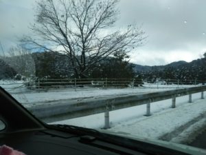
ということで向かった通天湖。道中は前日に降った雪で積雪もあり、釣り場も雪景色。
昼前からスタート。とりあえずキャストを教えるところから始まり、重めのスプーンで広く探りますがマスさんは口を使ってくれません。
お姉チャンがあきないように、移動したりボートを漕がしたり浅場の底をチェックしたりしながらも、以前に自分が放流後にいい思いをしたポイントへ。
これが大正解。1投目からフェザージグにレインボーが、2投目クランクにも。
さらに1.6gスプーンにも釣れてきて、どうも中層で浮いている感じ。
お姉チャンもテンション上がって投げるも、木に引っかかったりで、なかなか掛からず。
しかし、ホスト役に徹してコチラが巻いて止めてを教えると早々にヒット!
その後も掛かる→ばらすを何回か繰り返し、充分楽しんで時間となりました。
結果、お姉チャンも釣れて自分も満足した釣果に良い釣りができました。
「良かったなぁ釣れて。また付いて行ってあげるわ」
と帰りの車で、お褒めの言葉を頂きました。


