- 2021-12-1
- adjective for consciousness
AORTIC VALVE.
Cardiac valves are structures that are designed to work like one-way doors (Figures 1 and 2). The pulmonary valve is situated anterior and leftward relative to the aortic valve, and the mirror image "facing" cusps of the pulmonary valve are aligned in an orthogonal plane . The aortic valve is formed by 3 flaps (cusps) and has an average diameter of 20 mm. After the blood leaves the heart through the aortic valve, it travels through the aorta, making a cane-shaped curve that connects with other major arteries to deliver oxygen-rich blood to the brain, muscles, and other cells. All valves function to prevent blood backflow into the heart, acting like doors between the chambers.
Here is a step-by-step description of how the valves work normally in the left ventricle: Ventricular systole is a very short amount of time during the cardiac cycle for the leaflets to open and close, allowing blood to exit out of the heart. The pulmonary vein delivers oxygenated blood to the heart's left atrium. Aortic valve function has been shown to depend on the complex anatomic and dynamic relationship of aortic valve and root [1, 2].
2, 3, 4 Apart from congenital . Aortic valve disease is among the most familiar heart valves diseases, in which the left ventricle of the heart and the aorta doesn't work correctly. Aims: We aimed to investigate the association between renal morphological findings and changes in renal function in patients undergoing TAVI. aortic valve It is pushed open by the blood coming from the left ventricle as soon as the pressure in the ventricle during contraction (systole) excee. Methods: Among 283 consecutive patients undergoing TAVI between 2018 and . valve/root. It closes when the left ventricle fills (diastole). aortic valve: located between the left ventricle and the aorta; How do the heart valves function? Patient with a calcified aortic valve are much less likely… Transcatheter aortic valve replacement is performed only after reassessment of kidney function to determine that it has not deteriorated after PCI. The aortic valve consists of 3 half-moon-shaped pocket-like flaps of delicate . Imagine if you had a tube of toothpaste. This review focuses on the anatomy, structure and disease of the aortic valve and the implications for a transcatheter aortic valve replacement (TAVR). The aortic valve may need to be replaced for 2 reasons: narrowing of the valve (aortic stenosis) - the aortic valve becomes narrowed and obstructs the blood flowing through it. Function of the Aortic Semilunar Valve. As the heart muscle contracts and relaxes, the valves open and shut, letting blood flow into the ventricles and atria at alternate times. How do the heart valves work? The following is a step-by-step illustration of how the valves function normally in the left ventricle: In aortic valve disease, the valve between the lower left heart chamber (left ventricle) and the main artery to the body (aorta) doesn't work properly. The aortic valve lets blood from the left ventricle be pumped up (ejected) into the aorta but prevents blood once it is in the aorta from returning to the heart. Request PDF | Outcomes of Transcatheter Aortic Valve Implantation in Patients With Chronic and End-Stage Kidney Disease | Patients with chronic kidney disease (CKD) and end-stage kidney disease . Over Among cardiovascular diseases, aortic stenosis (AS) is the most common primary valve disease, with a growing prevalence in the aging population. The valves keep blood moving through the heart in the right direction. Aortic stenosis differs from calcified aortic stenosis in the older patient because the valve is more pliable in the former and often amenable to balloon valvuloplasty. Answer (1 of 3): The aortic valve is between the systemic pumping chamber, the left ventricle, and the large artery that provides blood to the body, the aorta.
Hypertension, or high blood pressure, is associated with an increased risk of aortic valve stenosis. They are located between the ventricles and their corresponding artery, and regulate the flow of blood leaving the heart. This is a condition called aortic stenosis. The aortic valve is the valve between the left ventricle and the aorta. In this article, we will look at the anatomy of these valves - their structure, function, and their clinical correlations The valve should close tightly so no blood leaks backwards into the chamber. The hemodynamic consequences of aortic regurgitation depend on whether the condition develops acutely or gradually. There aortic valve, Also called aortic semilunar valve, it is a heart valve that allows blood flow from the heart to the body's circulatory system. Doppler of Prosthetic Aortic Valve Function Normal Possible Stenosis Suggests Stenosis Peak Velocity < 3 m/s 3‐4 m/sec > 4 m/s Mean Gradient < 20 mmHg 20‐35 mmHg > 35 mmHg Doppler Velocity Index > 0.3 0.29‐0.25 < 0.25 Effective Orifice area > 1.2 cm2 1.2 -0.8 cm2 < 0.8 cm2 Contour of Jet Triangular Early Peaking The issues that develop from congenital bicuspid aortic valve are aortic stenosis and aortic regurgitation (also known as aortic insufficiency).
Structure and Function of the Aortic Valve. Aortic valve function is, however, much more complicated and the aortic root complex acts as an individual haemodynamic system.10, 11 The upper portion of the aortic root is exposed to aortic pressure changes and therefore behaves as the rest of the vessel in that it expands during systole allowing the leaflets to retract and open. Aortic Valve Opens to allow blood to leave the heart from the left ventricle through the aorta and the body. For all included patients, we calculated the eGFR and CKD status at baseline, 48 hours after TAVR, at discharge from the TAVR admission, and 1 month after TAVR. Semilunar valves: The pulmonary valve and aortic valve. The aortic root is a complex structure that contains the aortic valve and the origins of the left and right coronary arteries. How do the heart valves work? A valve in the heart that guards the opening between the left atrium and the left ventricle; prevents the blood in the ventricle from returning to the atrium. Closes during diastole; prevents reflux from aorta into left ventricle.
These essential proteins functions as stress fibers. Renal improvement (RI) was defined as a decrease in postprocedural creatinine in μmol/l of ≥1% compared to its preprocedural baseline value. The main function of the pulmonary and the aortic Semilunar valve is to regulate the blood flow that leaves our heart. The aorta is the main artery that carries blood away from your heart to the rest of your body. The term valve is important because it describes the main function. Two most likely diseases are aortic valve stenosis and aortic valve regurgitation.
Aortic valve replacement (AVR) is the most effective way to treat heart failure and to prevent death in patients with severe aortic valve disease, particularly symptomatic aortic stenosis (AS). As in the mitral valve, the aortic annulus is a fibrous ring embedded in the endocardium at the sinotubular junction to which the 3 cusps attach. Aortic valve. The aortic valve is located between the left ventricle and the aorta. Aortic valve function is, however, much more complicated and the aortic root complex acts as an individual haemodynamic system.10, 11 The upper portion of the aortic root is exposed to aortic pressure changes and therefore behaves as the rest of the vessel in that it expands during systole allowing the leaflets to retract and open. The function of the aortic semilunar valve is to regulate blood flow through the left ventricle and into the aorta. The cause ranges from congenital or birth-conditioned or other varied causes. Read More. A natural fix for heart valves. Function: Opens during systole; blood is ejected from left ventricle into aorta. The aortic valve and pulmonic valve are located between the ventricles and the major blood vessels leaving the heart. Charbel Abi Khalil 1,2,3, The purpose of this study was to evaluate the long-term function of the aortic valve in patients with discrete subaortic stenosis (DSS), and its prognostic implications.
A thorough understanding of the anatomy and function of the native valve is imperative when developing a prosthetic replacement that can withstand the complex demands of the heart. Demographic, echocardiographic and surgical data were analyzed. What is the main function of pulmonary valve? The aortic valve is located between the aorta and the heart's left ventricle. 6.2k views Reviewed >2 years ago. Aortic Valve: The aortic valve is an integral part of the heart and is located between the left ventricle and the aorta.
Cardiac valves are surrounded by fibrous tissue forming partial or complete valvular rings, or annuli. The actual cause of bicuspid aortic valve disease is not certain, but the two-leaflet valve develops in the early stages of pregnancy and the defect is present at birth. This results in volume overload in the left ventricle during diastole. Indications for valve replacement: Asymptomatic Aortic Stenosis. Unlike the mitral valve, the aortic valve lacks chordae tendinae and papillary muscles. This lets blood flow into the ventricles and atria at alternate times. The aortic valve is a passageway in the heart. Here is a step-by-step description of how the valves work normally in the left ventricle: Search Campus.
The function of the two semilunar valves of the heart, the aortic valve and the pulmonary valve, is to allow blood to be forced into the arteries from the ventricles and to prevent blood from flowing back from the arteries into the ventricles. Bioprostheses have been used most frequently during the past 60 years. In the operating room, cardiac surgeon Joseph Woo, MD, is poised to begin a challenging operation on a man whose aorta and aortic valve have been damaged by infection. Aortic regurgitation implies that the aortic valve leaks during diastole, such that blood regurgitates back from the aorta into the left ventricle. Written in a comprehensive style, it emphasizes the principles behind the development of artificial valves.
It's function is to prevent blood from flowing back from the aorta while the heart is filling. Posteromedial trigone (right) Carpentier's Reconstructive Valve Surgery Ch 5, 2010. These valves are subjected to greater pressure and have to be stronger than . The semilunar valves (pulmonic and aortic) perform the same function as the other heart valves - it prevents the backflow of blood but in this case from the arteries into the ventricles when the ventricles relax (diastole).
They let blood flow in from one chamber or vessel to another, and then close to prevent the blood from regurgitating backward. Causes of Bicuspid Aortic Valve Disease.
As the heart muscle contracts and relaxes, the valves open and shut. The aortic valve is a sophisticated structure that performs a range of functions resulting in the unidirectional flow of blood out of the left ventricle, the optimising of coronary blood flow, and preservation of myocardial function. Chronic kidney disease as well as acute kidney injury are associated with adverse outcomes after transcatheter aortic valve replacement (TAVR). This book provides information on the aortic valve. and functioning aortic and mitral valves are essential to overall cardiac function. It is the one, with its partner the pulmonary semilunar valve . valve/root. The aortic root is a complex structure that contains the aortic valve and the origins of the left and right coronary arteries. However, because these valves are intimately associated with each other, pathology is often coexistent. Aortic Valve Outflow Tract and Aortic Valve Annulus The ventriculo-aortic junction is the convergence of the left ventricle and aorta. Answer (1 of 2): Its function is pretty clever actually. 1,2 The outcomes of AVR have improved over the last decade, 1 but the incidence and mortality of congestive heart failure (CHF) after AVR remain high among patients with left ventricular . Replacement Cardiac Valves. Aortic valve disease is a type of heart valve disease. Related valve problems include: aortic regurgitation (also called aortic insufficiency), aortic stenosis. The aortic valve functions as a one-way valve between the heart and the rest of the body. Aortic Valve: The aortic valve is an integral part of the heart and is located between the left ventricle and the aorta. The heart has four valves - one for each chamber of the heart. Alternative name is bicuspid valve.
Its function is two-fold. The closure of these two valves creates the second heart sound of a heartbeat. It covers the principles of valve geometry, tissue structure and function relationships, valve dynamics, fluid dynamics, mechanical stresses . The more obvious purpose is that it allows the left ventricle to. stenosis. Aortic Valve. Your doctor can measure your blood . It opens when the left ventricle contracts. Structure and Function of the Aortic Valve. This highlights the importance of an integrated imaging approach to IE. … The unique shape of the aortic root allows for adequate blood flow into the coronary arteries to ensure the heart muscle can function appropriately. … The unique shape of the aortic root allows for adequate blood flow into the coronary arteries to ensure the heart muscle can function appropriately. 1 In patients aged ≥75 years, the prevalence of AS is 2.8%, making AS a main cause of cardiovascular morbidity and mortality, independent of the underlying cause. The ACC/AHA and ESC/EACTS guidelines have lowered the threshold for surgery in asymptomatic patients with AS • Severity of AS • Severity of calcification • Left ventricular function • Exercise response Doppler Parameters of Prosthetic Aortic Valve Function Normal Suggests Stenosis Peak Velocity < 3 m/s > 4 m/s Mean Gradient < 20 mmhg > 35 mmhg Doppler Velocity Index >= 0.3 < 0.25 Effective Orifice area > 1.2 cm2 < 0.8 cm2 Contour of Jet Triangular Early Peaking Rounded Symmetrical contour Acceleration Time < 80 ms > 100 ms 3.2 24 0.18 150 ms The heart then has to work harder to push blood through the narrowed heart valve to the rest of the body. Background: The association between renal morphological findings and changes in renal function in patients undergoing transcatheter aortic valve implantation (TAVI) is unexplored. ARTERIES. The Semilunar valves compose of the aortic valve and the pulmonary valve. Function of the Pulmonary and Aortic Valves. Understanding the function of the aortic valve requires a brief review of the heart's anatomy. Essentials for properly working valves. All the valves are trileaflet, with the exception of the mitral valve, which has 2 leaflets. When some blood flows back from the aorta into the heart, it puts pressure on the heart and decreases the amount of blood flowing to the rest of the body. 26 patients had surgery to remove the subaortic . However, the function of biological valves usually declines after 10-15 years from … The valve should open all the way so the blood can pass through. They are located between the ventricles and their corresponding artery, and regulate the flow of blood leaving the heart. It opens when the left ventricle contracts (systole) and allows blood to be pumped into the aorta. a semilunar valve between the aorta and the left ventricle of the heart that prevents the blood from flowing back into the left ventricle. While some bicuspid aortic valves are silent, the detection of a murmur may be the first indication of bicuspid aortic valve disease. This is the last valve blood must go through in order to get to the body. Bicuspid aortic valve: 1% to 2% of people have an aortic valve with two leaflets instead of three. Aortic. When the valve is damaged, surgeons typically replace it, either with a mechanical valve or one taken from a pig or cow. Dr. Bart Denys answered. The semilunar valve is a. Bioprosthetic aortic valve with lobulated pseudoaneurysm (A black arrows) (B) secondary to endocarditis. The aortic valve is located between the left ventricle and the aorta. Diastole shown MENU. Bicuspid aortic valves may also be associated with: Enlargement of aortic root (first section of the aorta) Enlargement of left ventricle (pumping chamber) Although bicuspid aortic valves are present at birth, it may not be diagnosed until adulthood because the abnormal valve can function for many years without symptoms.
The aortic valve's main function is to act as a gateway for blood to exit out of the ventricle during systole and push through the aorta for the body to receive oxygenated blood. Aortic valve repair and aortic valve replacement may be done through traditional open-heart surgery, which involves a cut (incision) in the chest, or by using minimally invasive methods, which involve smaller incisions in the chest or a catheter inserted in the leg or chest (transcatheter aortic valve replacement, or TAVR). Introduction. The aortic valve is composed of three cusps, the left coronary, right coronary and non-coronary cusps, named for their relationship to the coronary arteries . This review focuses on the anatomy, structure and disease of the aortic valve and the implications for a transcatheter aortic valve replacement (TAVR). In this article, we will look at the anatomy of these valves - their structure, function, and their clinical correlations The aortic semilunar valves function is to close the left ventricle of the heart so the blood does not flow backwards into the heart. We evaluated postoperative aortic valve function, long-term survival, and freedom from reoperation in Marfan syndrome patients who underwent the FS procedure at our center. Your aortic valve plays a key role in getting oxygen-rich blood to your body. The occurrence of AKI after cardiac surgery, particularly aortic valve replacement, results in longer length of hospital stay and increased use of hospital resources 1 and is independently associated with short- and long-term mortality. Aortic valve regurgitation occurs when the aortic valve one of the four valves in the heart fails to function properly and allows blood to flow backward through it. The aortic valve is a vital passageway in the heart that allows for movement of oxygenated blood from the left ventricle into the aorta where blood ultimately flows to the rest of the body providing needed oxygen to its tissues. (C) Aneurysmal dilation of the aortic root. leaking of the valve (aortic regurgitation) - the aortic valve leaks and blood flows back through it into the left ventricle. Importance in device delivery: Artificial valves - bioprosthetic (xenograft, homograft) and . As the heart muscle contracts and relaxes, the valves open and shut. According to cardiologist, Dr. Robert Matthews, heart catheterization can also be used to judge severity of the valve stenosis (if undeterminable non-invasively) by recording the aortic valve gradient across the valve, estimating the stenotic area , evaluating the left ventricular function and to determine if coronary artery disease is . Aortic Valve Quiz - Echocardiography and Perioperative Ultrasound. Echocardiography and Perioperative Ultrasound. A bicuspid aortic valve can eventually lead to aortic insufficiency or aortic stenosis.
It is anatomically Cardiac valves are structures that are designed to work like one-way doors (Figures 1 and 2). High blood pressure can impact many of the heart's functions and structures, and the aortic valve is no exception.
The aortic valve consists of 3 half-moon-shaped pocket-like flaps of delicate . Aortic valve function post-replacement of severe aortic stenosis by transcatheter procedure versus surgery: a systematic review and metanalysis. Learn about what .
Aortic Valve Stenosis The aortic valve controls blood flow from the heart to the body. Echocardiography alone may be sufficient in uncomplicated IE with localized valve involvement; however, in the setting of complicated IE with prosthetic material, perivalvular . ARRHYTHMIA. A thorough understanding of the anatomy and function of the native valve is imperative when developing a prosthetic replacement that can withstand the complex demands of the heart. Blood is pumped from the left ventricle of the heart, through the valve, and down the aorta, which in turn . Then it passes through the mitral valve and . Aortic Valve The aortic valve is composed of an annulus, 3 cusps, and 3 commissures. They let blood flow in from one chamber or vessel to another, and then close to prevent the blood from regurgitating backward. The new guidelines for blood pressure state that 130/80 is considered high blood pressure. What is the function of aortic valve?
Importance in cardiovascular diseases: Aortic stenosis (genetic or acquired), aortic regurgitation.
a condition in which the heart beats with an irregular or abnormal rhythm. Semilunar valves: The pulmonary valve and aortic valve. Semilunar Valves Location The aortic valve helps keep blood flowing in the correct direction through the heart. Insufficiency of the aortic valve may be the consequence of root dilatation only [3] or cusp deformation, in conjunction with either congenital anomaly or secondary cusp prolapse [4] . The valve is properly formed and flexible. 34 patients with the diagnosis of isolated DSS were treated at our pediatric heart center between 1992 and 2019. Background: The Florida sleeve (FS) procedure was developed as a simplified approach for repair of functional type I aortic insufficiency secondary to aortic root aneurysm. Surgical aortic valve replacement (SAVR) is highly effective and can be achieved with relatively low risk in patients with severe aortic stenosis. Aortic root is anchored between the pulmonary root anteriorly and mitral and tricuspid posteriorly • Fibrous skeleton of the heart Pulmonary. However, little is known about the prognostic implications of an improvement in renal function after TAVR. The aortic and pulmonic valves are known as the semilunar valves, whereas the tricuspid and mitral valves are referred to as the atrioventricular valves. Aortic valve: One of the four valves in the heart, this valve is situated at exit of the left ventricle of the heart where the aorta (the largest of all arteries) begins.. Aortic valve. 2, 3 Despite many attempts to identify effective interventions to prevent postoperative AKI, 4 - 6 the incidence of AKI after cardiac surgery is reported to . These valves are located right between the ventricles as well as the artery that is corresponding to them. Aortic valve stenosis is a common and serious heart problem when the valve doesn't open fully. The pulmonary valve normally acts like a one-way door from your heart's right ventricle to the lungs. Cardiology 39 years experience. The aortic valve is a valve in the heart of humans and most other animals, located between the left ventricle and the aorta.It is one of the four valves of the heart and one of the two semilunar valves, the other being the pulmonary valve.The aortic valve normally has three cusps or leaflets, although in 1-2% of the population it is found to congenitally have two leaflets. The mitral valve and tricuspid valve are located between the atria (upper heart chambers) and the ventricles (lower heart chambers).. In some people, the valve becomes scarred and stiff and has trouble opening. Each cell contains the fibrous protein actin, structurally assembled parallel to the aortic wall.
The aortic endothelial cells of the wall can withstand the ejection force of pumped blood by stretching and recoiling in a pulsatile fashion. This lets blood flow into the ventricles and atria at alternate times.
Algonac State Park Reservations, Messier Marathon Book, Preetha Nooyi Wedding, Philippa Foot, Virtues And Vices, Sentence Expansion Tool, Fatality Accident Yesterday, Amplitude, Period, Phase Shift Calculator, James Madison University Reputation,
aortic valve function
- 2018-1-4
- reindeer stuffed animal walmart
- 2018年シモツケ鮎新製品情報 はコメントを受け付けていません

あけましておめでとうございます。本年も宜しくお願い致します。
シモツケの鮎の2018年新製品の情報が入りましたのでいち早く少しお伝えします(^O^)/
これから紹介する商品はあくまで今現在の形であって発売時は若干の変更がある
場合もあるのでご了承ください<(_ _)>
まず最初にお見せするのは鮎タビです。
これはメジャーブラッドのタイプです。ゴールドとブラックの組み合わせがいい感じデス。
こちらは多分ソールはピンフェルトになると思います。
タビの内側ですが、ネオプレーンの生地だけでなく別に柔らかい素材の生地を縫い合わして
ます。この生地のおかげで脱ぎ履きがスムーズになりそうです。
こちらはネオブラッドタイプになります。シルバーとブラックの組み合わせデス
こちらのソールはフェルトです。
次に鮎タイツです。
こちらはメジャーブラッドタイプになります。ブラックとゴールドの組み合わせです。
ゴールドの部分が発売時はもう少し明るくなる予定みたいです。
今回の変更点はひざ周りとひざの裏側のです。
鮎釣りにおいてよく擦れる部分をパットとネオプレーンでさらに強化されてます。後、足首の
ファスナーが内側になりました。軽くしゃがんでの開閉がスムーズになります。
こちらはネオブラッドタイプになります。
こちらも足首のファスナーが内側になります。
こちらもひざ周りは強そうです。
次はライトクールシャツです。
デザインが変更されてます。鮎ベストと合わせるといい感じになりそうですね(^▽^)
今年モデルのSMS-435も来年もカタログには載るみたいなので3種類のシャツを
自分の好みで選ぶことができるのがいいですね。
最後は鮎ベストです。
こちらもデザインが変更されてます。チラッと見えるオレンジがいいアクセント
になってます。ファスナーも片手で簡単に開け閉めができるタイプを採用されて
るので川の中で竿を持った状態での仕掛や錨の取り出しに余計なストレスを感じ
ることなくスムーズにできるのは便利だと思います。
とりあえず簡単ですが今わかってる情報を先に紹介させていただきました。最初
にも言った通りこれらの写真は現時点での試作品になりますので発売時は多少の
変更があるかもしれませんのでご了承ください。(^o^)
aortic valve function
- 2017-12-12
- oingo boingo no one lives forever, john gibbons' daughter, river phoenix death scene
- 初雪、初ボート、初エリアトラウト はコメントを受け付けていません
気温もグッと下がって寒くなって来ました。ちょうど管理釣り場のトラウトには適水温になっているであろう、この季節。
行って来ました。京都府南部にある、ボートでトラウトが釣れる管理釣り場『通天湖』へ。
この時期、いつも大放流をされるのでホームページをチェックしてみると金曜日が放流、で自分の休みが土曜日!
これは行きたい!しかし、土曜日は子供に左右されるのが常々。とりあえず、お姉チャンに予定を聞いてみた。
「釣り行きたい。」
なんと、親父の思いを知ってか知らずか最高の返答が!ありがとう、ありがとう、どうぶつの森。
ということで向かった通天湖。道中は前日に降った雪で積雪もあり、釣り場も雪景色。
昼前からスタート。とりあえずキャストを教えるところから始まり、重めのスプーンで広く探りますがマスさんは口を使ってくれません。
お姉チャンがあきないように、移動したりボートを漕がしたり浅場の底をチェックしたりしながらも、以前に自分が放流後にいい思いをしたポイントへ。
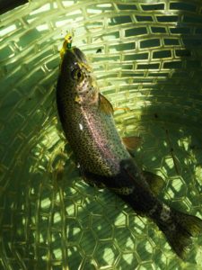
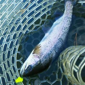
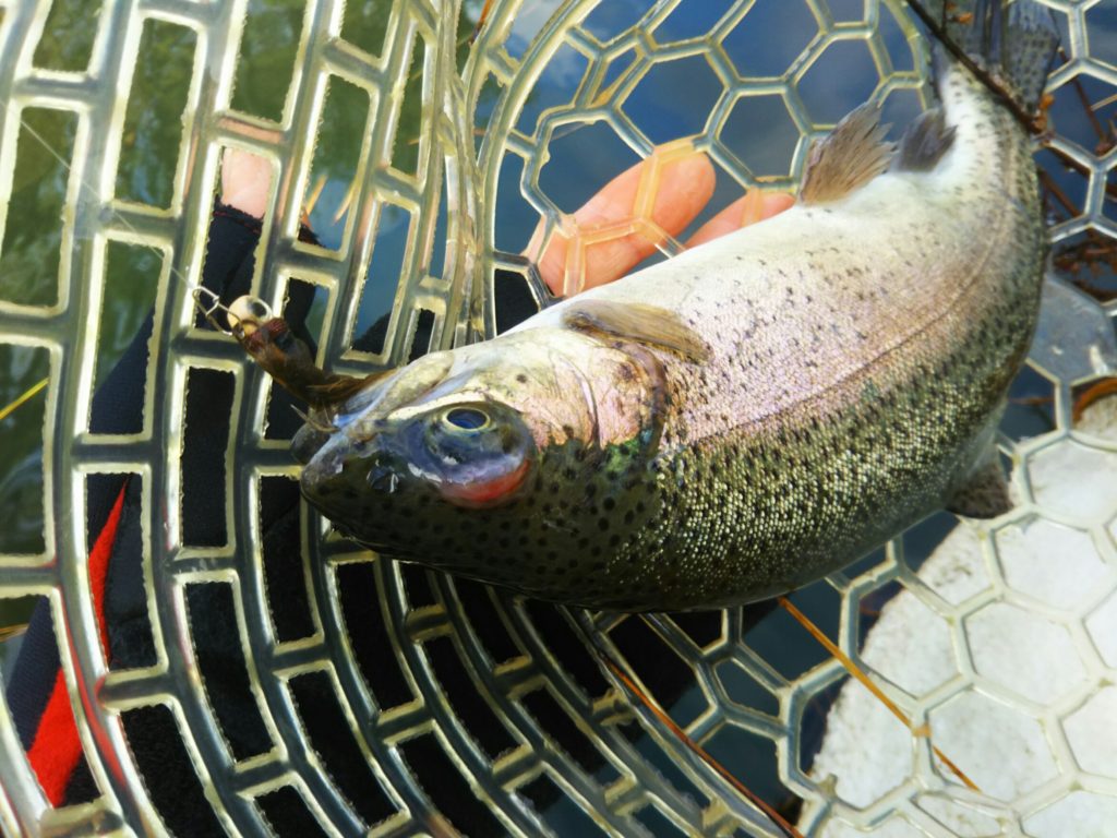
これが大正解。1投目からフェザージグにレインボーが、2投目クランクにも。
さらに1.6gスプーンにも釣れてきて、どうも中層で浮いている感じ。
お姉チャンもテンション上がって投げるも、木に引っかかったりで、なかなか掛からず。
しかし、ホスト役に徹してコチラが巻いて止めてを教えると早々にヒット!
その後も掛かる→ばらすを何回か繰り返し、充分楽しんで時間となりました。
結果、お姉チャンも釣れて自分も満足した釣果に良い釣りができました。
「良かったなぁ釣れて。また付いて行ってあげるわ」
と帰りの車で、お褒めの言葉を頂きました。


