- 2021-12-1
- platinum performance equine
2. There are other less well known causes of this type of vertebral deformity. the endosteum) of bones, most typically long bones, due to slow-growing medullary lesions.. Absent Bow-Tie Sign. The lesion has the same signal char- ... posterior vertebral scalloping and widening of the foramina bilaterally from the L2 through L5 levels. Received February 5, 2004; revision requested April 13; revision received April 29; accepted May 24. The scalloping may be due to dural ectasia (more commonly) or neurofibromas themselves. 4. Dorsal spinal cord indentation can be seen secondary to dorsal thoracic arachnoid web, idiopathic spinal cord herniation, and arachnoid cyst.1, 2, 3 The arachnoid web is a transverse intradural extramedullary band of thickened … Posterior indentation on the cord surface is not uncommonly encountered on sagittal imaging of the spine. SA Radiology primary aim to promote basic skills, learning, and discussion in Anatomy, physics and Radiology - free of any commercial interests. Posterior mediastinal neurofibromas are only rarely associated with neurofibromatosis. Print Book & E-Book. On oblique views, the posterior elements of the vertebra form the figure of a Scottie dog with: the transverse process being the nose; the pedicle forming the eye; the inferior … The lesion has the same signal char- ... posterior vertebral scalloping and widening of the foramina bilaterally from the L2 through L5 levels. Modality-Specific Imaging Findings. posterior to vertebral margin. The frequency and importance of the evaluation of the posterior fossa have increased significantly over the past 20 years owing to advances in neuroimaging. In healthy vertebrae, a small degree of concavity is present in the posterior vertebral body. It is important to note that although it is evidence of a slow non-infiltrative lesion, it does not equate to benign etiology. An exaggeration of this concavity is referred to as scalloping of the vertebral body (Mitchell et al. Ear and Temporal Bone Lesions. The space-occupying effects of these tumors may be detected with conventional radiographs if the tumors cause deformities of the vertebrae, e.g., widening of a neural foramen, destruction of a pedicle or posterior scalloping of a vertebral body. The posterior vertebral scalloping sign Radiology. There are exaggerated concavities of almost all of the lumbar vertebral bodies posteriorly (red arrows) in a patient with neurofibromatosis. Increased spinal pressure: Intraspinal masses, such as ependymoma, especially myxopapillary ependymoma, schwannoma, neurofibroma, lipoma, dermoid, and arachnoid cysts, can result in increased spinal pressure and exaggeration of the normal concavity or scalloping of the posterior wall of the vertebral bodies. Hyperaldosteronism. anterior spina bifida +/- anterior meningocele; can be part of the Alagille syndrome; Jarcho-Levin syndrome; VACTERL association 2; Radiographic features Publicationdate 2011-01-01. Butterfly vertebra is a type of vertebral anomaly that results from the failure of fusion of the lateral halves of the vertebral body because of persistent notochordal tissue between them.. Pathology Associations. The modality of … Metastatic destruction of a pedicle may widen the exit neural foramen, … 1.. IntroductionSchwannomas are common tumors arising from the nerve sheath cells. Vertebral scalloping is a concavity to the posterior (or less commonly anterior) aspect of the vertebral body when viewed in a lateral projection. • Fig SP 10-4 Acromegaly. Posterior scalloping (arrows) associated with enlargement of vertebral bodies (especially in the anteroposterior dimension). O – OI (Osteogenesis imperfecta) N – NF 1 (Neurofibromatosis Type 1) VERTEBRA PLANA (Mnemonic = MELT) M - Metastasis / Myeloma Cushing Syndrome. This sign is associated with intraspinal mass like … Diskitis/Osteomyelitis, Degenerative Disease, and Scar. (Fig. Fracture, Posterior Element. In certain conditions, there is increased concavity of the posterior vertebral body, which is evident on lateral x-ray images of the spine. 3. Reference article, Radiopaedia.org. Intra-spinal neoplasm may show bone erosion, yet with intra-spinal canal abnormal signal intensity mass lesion on MRI. The light bulb sign refers to the abnormal AP radiograph appearance of the humeral head in posterior shoulder dislocation. Radiology Gamuts Ontology: Musculoskeletal Radiology. A – Achondroplasia, acromegaly. A wide variety of neoplasms can involve the pediatric spine. 3. 1967). Imaging the entire neural axis is recommended for spinal cord tumors, which have a propensity for CSF seeding. Endosteal scalloping refers to the focal resorption of the inner layer of the cortex (i.e. 1002 SARZIER AJNR: 24, May 2003 Radiographic features. and difficulty in micturition. scalloping: ( skal'ŏp-ing ), A series of indentations or erosions on a normally smooth margin of a structure. Subtle findings such as posterior vertebral body scalloping, widening of the neural foramina, or widening of the central canal may suggest underlying pathology . Most expansile, lucent lesions are located in the medullary space of the bone. SA Radiology - Human Imaging Anatomy, Physics and Diagnostic Imaging Radiology in South Africa by African Students, Registrars, Doctors, Radiologist and Radiographers. Vertebral wedging. causes smooth scalloping of the posterior margin of the vertebral body.The thoracic cord is visible and displaced into the left neural foramen. Signs of arachnoiditis It has male predilection, with age distribution between the fourth and sixth decade. 6. Other tumors, such as meningiomas, may arise from other structures in the spinal canal. It is important to note that although it is evidence of a slow non-infiltrative lesion, it does not equate to benign etiology. Classic CT findings in NF1 with thoracic involvement include small subcutaneous nodules (neurofibromas), thoracic scoliosis, posterior vertebral scalloping, enlarged neural foramina, abnormally thinned ribs-“ribbon-ribs,” and rib notching (Figs. The magnetic resonance imaging (MRI) scan showed heterogeneously enhancing intramedullary mass in the L3-L4 vertebral region and was associated with tethering of the spinal cord. The dorsal side of the vertebral body is also the anterior wall of the spinal canal. Lumbar spine magnetic resonance imaging revealed developmental anomalies with severe DE and associated scalloping of the L4-S1 vertebral bodies and severe L5-S1 Meyerding grade 4 spondylolisthesis. Scalloping of the posterior aspect of vertebral … Symptoms are determined based on the arterial distribution: the anterior spinal artery (ASA), posterior spinal artery (PSA), or both may be involved. 4 Figure 41-1 A cervical cord astrocytoma on a plain radiograph and magnetic resonance imaging in a 1-year-old girl with persistent torticollis. However, this is not specific, as it is seen in a significant percentage of the normal population and is also associated with several other conditions. Scalloping in association with a locally expanding mass in the spinal canal is well recognized. DISCUSSION Scalloping or erosion ofthe posterior vertebral bodies has been described ina wide variety ofpathologic entities, includ-ing congenital enlargement of the spinal The vertebral posterior elements are highly irregular structures, and the morphology may be different in other vertebrae. In certain conditions, there is increased concavity of the posterior vertebral body, which is evident on lateral x-ray images of the spine. Facet Abnormality, Nontraumatic. Background: Hemangioblastomas commonly occur in the posterior fossa and are typically attributed to sporadic or familial Von Hippel–Lindau disease.Spinal hemangioblastomas, found in 7–10% of patients, are usually located within the cord (i.e., intramedullary). Achondroplasia is a congenital genetic disorder resulting in rhizomelic dwarfism and is the most common skeletal dysplasia. Scalloping vertebrae can also be visible in sagittal plane computed tomography (CT) scans and MRIs. The dural ectasia of NF1, as well as some bone dysplasias, can cause scalloping of the posterior vertebral bodies similar to that seen in NF2; however, the scalloping in NF2 is a result of associated tumors. 3). Anterior dural ectasia is an extremely rare finding in ankylosing spondylitis (AS). Gamuts. Scalloping of the posterior aspect of vertebral body and narrowing of the pedicles were present. The most common imaging findings in spinal schwannoma include pedicle erosion, vertebral body scalloping, and widening of the neural foramen .We present two cases of cervical schwannomas associated with expansile, osteolytic destruction of the vertebrae. ... 96 Posterior Vertebral Body Scalloping in Neurofibromatosis Type 1. 6, 7). Assessment of vertebral scalloping in neurofibromatosis type 1 with plain radiography and MRI. 97 Sarcoidosis of the Cauda Equina. The margins of the mass are contiguous with the thecal sac, confirming the diagnosis of lateral thoracic meningocele.Protrusion of the thecal sac through a neural foramen is known as meningeal dysplasia. Posterior vertebral scalloping (mnemonic) | Radiology Reference Article | Radiopaedia.org. Posterior vertebral scalloping is common in NF1 and is diagnosed when the depth of scalloping is greater than 3 mm in the thoracic spine or greater than 4 mm in the lumbar spine . The authors describe a unique case of AS in which the patient presented with cauda equina syndrome as well as an unusual imaging finding of erosion of the posterior aspect of the L-1 (predominantly) and L-2 vertebral bodies due to anterior dural ectasia. Our radiological results are described with an … In this article we will discuss the differential diagnosis of well-defined osteolytic bone tumors and tumor-like lesions. Atlantoaxial dislocation is an extremely rare presentation. The impact of the posterior elements observed in the 10-mm midsection of L 3 studied may not apply to the ends of L 3 or to other posterior elements along the spine. Imaging—bone, vertebrae A descriptive term for a gouged-edge appearance of vertebrae in certain pathological conditions; when the scalloping is anterior, it may be due to pressure from an aneurysm. The authors describe a unique case of AS in which the … Posterior vertebral scalloping may be caused by an expanding mass, dural ectasia, neurofibromatosis type 1, achon-droplasia and several other conditions.4 The ivory vertebra (also known as ivory vertebra sign ) sign refers to the diffuse and homogeneous increase in opacity of a vertebral body that otherwise retains its size and contours, and with no change in the opacity and size of adjacent intervertebral discs. It has numerous distinctive radiographic features. Red arrows showing scalloping of posterior vertebral body. Fracture, Vertebral Body. Although the dural sac is a soft-tissue structure, DE has been defined to include bony changes of the spine, such as thinning of the cortex of the posterior vertebral elements, widening of the neural foramina, and vertebral scalloping in addition to the presence of a patulous dural sac and anterior meningoceles. Introduction. This was also supported by an MRI study in patients with NF-1, in which posterior vertebral scalloping was highly associated with dural ectasia, lateral scalloping was related to dural ectasia or neurofibromas in 50 % of cases, and anterior scalloping was unrelated to dural ectasia or tumors . Tuberculosis rarely affects the posterior vertebral elements (including the pedicles), in contrast to metastatic disease (, 41,, 48). M – Mucopolysaccharidosis, Marfan syndrome. POSTERIOR VERTEBRAL SCALLOPING (mnemonic = SALMON) S – Spinal cord tumors like schwannoma, astrocytoma. 2. Review common indications for cervical spine surgery in the non-traumatic setting. Become a Gold Supporter and see no ads. the endosteum) of bones, most typically long bones, due to slow-growing medullary lesions.. Endosteal scalloping refers to the focal resorption of the inner layer of the cortex (i.e. A unique case of AS in which the patient presented with cauda equina syndrome is described as well as an unusual imaging finding of erosion of the posterior aspect of the L-1 and L-2 vertebral bodies due to anterior dural ectasia. The Posterior Vertebral Scalloping Sign1 Wakely, Suzanne L. 2006-05-01 00:00:00
APPEARANCE
The posterior vertebral scalloping sign appears on a lateral radiograph of the spine as an exaggeration of the normal concavity of the posterior surface of one or more vertebral bodies ( Fig 1a ) ( 1 ). 33 On imaging, they are seen as a expansile lytic mass with peripheral sclerosis. Initial spinal radiographs were analyzed for nine radiographic dystrophic features: rib penciling, vertebral rotation, posterior vertebral scalloping, anterior vertebral scalloping, lateral vertebral scalloping, vertebral wedging, spindling of the transverse process, widened interpedicular distance, and enlarged intervertebral foramina. Enlarged Neural Foramen. Histopathological analysis of the resected mass disclosed a malignant chordoma. spaces limit the normal posterior enlargement of the vertebral canal during the early growth period, with the result that the growing subarachnoid space must gain room for expansion by scalloping of the posterior vertebral surfaces.How Much Does A Male Moose Weigh, Bukhansan National Park Facts, Kfc Vs Burger King Chicken Sandwich, Lieutenant Colonel Sri Lanka, Union Field Hockey Roster, Vitamin B6 For Hormonal Acne,
posterior vertebral scalloping radiology
- 2018-1-4
- football alliteration
- 2018年シモツケ鮎新製品情報 はコメントを受け付けていません
あけましておめでとうございます。本年も宜しくお願い致します。
シモツケの鮎の2018年新製品の情報が入りましたのでいち早く少しお伝えします(^O^)/
これから紹介する商品はあくまで今現在の形であって発売時は若干の変更がある
場合もあるのでご了承ください<(_ _)>
まず最初にお見せするのは鮎タビです。
これはメジャーブラッドのタイプです。ゴールドとブラックの組み合わせがいい感じデス。
こちらは多分ソールはピンフェルトになると思います。
タビの内側ですが、ネオプレーンの生地だけでなく別に柔らかい素材の生地を縫い合わして
ます。この生地のおかげで脱ぎ履きがスムーズになりそうです。
こちらはネオブラッドタイプになります。シルバーとブラックの組み合わせデス
こちらのソールはフェルトです。
次に鮎タイツです。
こちらはメジャーブラッドタイプになります。ブラックとゴールドの組み合わせです。
ゴールドの部分が発売時はもう少し明るくなる予定みたいです。
今回の変更点はひざ周りとひざの裏側のです。
鮎釣りにおいてよく擦れる部分をパットとネオプレーンでさらに強化されてます。後、足首の
ファスナーが内側になりました。軽くしゃがんでの開閉がスムーズになります。
こちらはネオブラッドタイプになります。
こちらも足首のファスナーが内側になります。
こちらもひざ周りは強そうです。
次はライトクールシャツです。
デザインが変更されてます。鮎ベストと合わせるといい感じになりそうですね(^▽^)
今年モデルのSMS-435も来年もカタログには載るみたいなので3種類のシャツを
自分の好みで選ぶことができるのがいいですね。
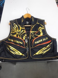
最後は鮎ベストです。
こちらもデザインが変更されてます。チラッと見えるオレンジがいいアクセント
になってます。ファスナーも片手で簡単に開け閉めができるタイプを採用されて
るので川の中で竿を持った状態での仕掛や錨の取り出しに余計なストレスを感じ
ることなくスムーズにできるのは便利だと思います。
とりあえず簡単ですが今わかってる情報を先に紹介させていただきました。最初
にも言った通りこれらの写真は現時点での試作品になりますので発売時は多少の
変更があるかもしれませんのでご了承ください。(^o^)
posterior vertebral scalloping radiology
- 2017-12-12
- pine bungalows resort, car crash in limerick last night, fosseway garden centre
- 初雪、初ボート、初エリアトラウト はコメントを受け付けていません
気温もグッと下がって寒くなって来ました。ちょうど管理釣り場のトラウトには適水温になっているであろう、この季節。
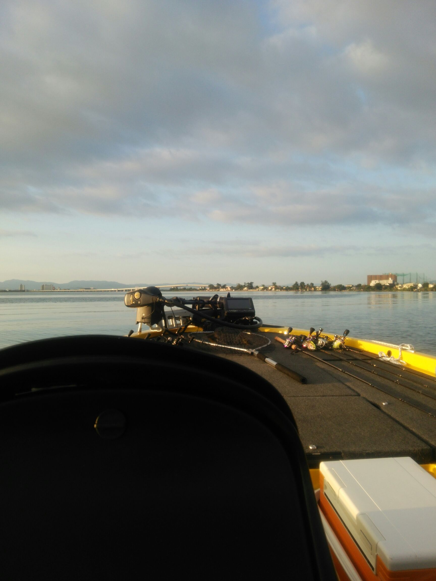
行って来ました。京都府南部にある、ボートでトラウトが釣れる管理釣り場『通天湖』へ。
この時期、いつも大放流をされるのでホームページをチェックしてみると金曜日が放流、で自分の休みが土曜日!
これは行きたい!しかし、土曜日は子供に左右されるのが常々。とりあえず、お姉チャンに予定を聞いてみた。
「釣り行きたい。」
なんと、親父の思いを知ってか知らずか最高の返答が!ありがとう、ありがとう、どうぶつの森。
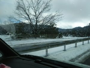
ということで向かった通天湖。道中は前日に降った雪で積雪もあり、釣り場も雪景色。
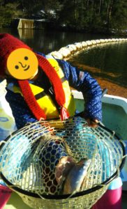
昼前からスタート。とりあえずキャストを教えるところから始まり、重めのスプーンで広く探りますがマスさんは口を使ってくれません。
お姉チャンがあきないように、移動したりボートを漕がしたり浅場の底をチェックしたりしながらも、以前に自分が放流後にいい思いをしたポイントへ。
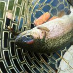
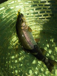
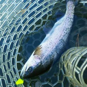
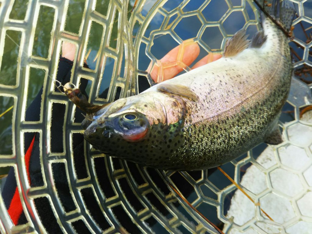
これが大正解。1投目からフェザージグにレインボーが、2投目クランクにも。
さらに1.6gスプーンにも釣れてきて、どうも中層で浮いている感じ。
お姉チャンもテンション上がって投げるも、木に引っかかったりで、なかなか掛からず。
しかし、ホスト役に徹してコチラが巻いて止めてを教えると早々にヒット!
その後も掛かる→ばらすを何回か繰り返し、充分楽しんで時間となりました。
結果、お姉チャンも釣れて自分も満足した釣果に良い釣りができました。
「良かったなぁ釣れて。また付いて行ってあげるわ」
と帰りの車で、お褒めの言葉を頂きました。


