- 2021-12-1
- platinum performance equine
Symptoms of renal colic and gross hematuria, pathognomonic of urolithiasis in adults, are seen less reliably in children. It is often caused by acute obstruction of the urinary tract by a calculus and is frequently associated with nausea and vomiting. Classical renal colic pain is located in the costovertebral angle, lateral to the sacrospinus muscle and beneath the 12th rib. Point-of-care (POC) renal ultrasound (US) is a rapid, bedside test for the evaluation of the patient with suspected renal colic or urinary retention. Your baby's doctor will do a complete physical exam to identify any possible causes for your baby's distress. Examining the limbs, fingers, toes, eyes, ears and genitals. For intermediate patients, ultrasound is a great starting point. Usual presentation of renal/ureteric stones is as an acute episode with severe pain (1) - renal colic or ureteric colic although some stones are picked up incidentally during imaging or may present as a history of infection the initial diagnosis is made by taking a clinical history and examination and carrying out imaging; initial management is . 3. Renal colic is a common reason for presentation to emergency departments, and imaging has become fundamental for the diagnosis and clinical management of this condition. Most of the patients (58.5%) had an age between 19 and 39 years, and the majority (60.1%) were males and had a body mass index (BMI) above 25. In one series of 134 patients with symptomatic AAA presenting to the ED, the following statistics were reported [ 23 ] : Eighteen percent had an initial misdiagnosis of nephrolithiasis. 2016 2017 2018 2019 2020 2021 2022 Billable/Specific Code. The pain of a leaking AAA is often misdiagnosed initially as renal colic. Ultrasonography and particularly noncontrast computed tomography have good diagnostic performance in diagnosing renal colic. Once the pain subsides, treatment is focused on the cause that has triggered renal . 1. Ureters carry urine from your kidneys to your bladder. Acute kidney injury is a concern in patients with . Risk factors for DVT and renal colic should be elicited during the history, as these can help narrow the diagnosis. Radiologic management will depend on the tools . This review will focus on calculus-related renal or ureteric coli … Calculi within the kidney do not cause pain. Receiver operating characteristic (ROC) curves for US diagnosis of acute urinary tract . The prevalence of renal colic was 0.5%. Common symptoms of renal colic. Incidence same as non-pregnant population. Infection) from a Kidney Stone: Fever strongly suggests Pyelo. Renal colic in pregnancy. 4 Noncontrast computed tomography in obstructive anuria: a prospective study • Pain from these structures is felt in these dermatomes. Variation in practice surrounding the diagnosis and management of suspected renal colic could have substantial implications for both quality and cost of care as well as patient radiation burden. Kidney Stone pain radiates to groin / genitals. Multiple risk factors include chronic dehydration. Let us have a look at the common symptom of renal colic: Renal colic pains come and go. These patients are concerning for either . The symptoms of renal colic vary depending on the size of the stone and its location in the urinary tract. Part 2: Diagnosis. I will reiterate my initial thought: imaging for renal colic is pretty easy. A prospective study of 98 patients presenting with acute flank or abdominal pain or both was conducted to determine the diagnostic accuracy of ultrasound scan compared with excretory urography for the diagnosis of urinary tract calculi. Unspecified renal colic. Like the non-pregnant person, 70-80% of the symptomatic stones pass spontaneously. P = Adult ED patients with signs/symptoms of renal colic I = IV Lidocaine (1.5 mg/kg) with or without IV Morphine (0.1 mg/kg) C = placebo with or without IV Morphine (0.1mg/kg) O = Pain, nausea, side effects Background Renal colic affects 1.2 million people and accounts for 1% of ED visits, with symptom control Receiver operating characteristic (ROC) curves for US diagnosis of acute urinary tract . The pain from kidney colic is very severe and can be extremely frightening for the patient, but its treatment is simple and its appearance can be avoided with some changes in habits. Previous literature has suggested that CT scanning has increased with no improvements in outcome . Renal colic diagnosis Diagnosis is made through a combination of history and physical exam, laboratory and imaging studies. Acute renal colic is a severe form of sudden flank pain that typically originates over the costovertebral angle and extends anteriorly and inferiorly towards the groin or testicle. - After 6 minutes you will be asked a series of questions by the examiner. IntroductionIntroduction • T10-12, L1 roots innervate the renal capsule and ureter. All patients underwent standardized ultrasound scan and . Renal colic, which is usually associated with kidney stones, is an urgent urologic condition that presents with severe, acute pain and is usually diagnosed and treated in the . Renal colic or kidney colic is a common condition that sends hundreds of patients to hospital every year. Other signs and symptoms that may occur with renal colic: Severe low back, abdominal, or groin pain Renal colic due to kidney stone develops suddenly with excruciating pain in back, side and radiates to the groin. The characteristic of any pain that's called 'colic' is in its spasmodic . Care map information Scope: • diagnosis and management of renal colic in primary care in adults, including pregnant women Renal colic is a common reason for presentation to emergency departments, and imaging has become fundamental for the diagnosis and clinical management of this condition. The degree of pain is related to the degree of obstruction and not the size of the . Urine is dark colored, there may be blood in urine, fever, chill, vomiting are other complimentary symptoms. For claims with a date of service on or after October 1, 2015, use an equivalent ICD-10-CM code (or codes). Signs and symptoms. blood in your urine, which may be pink, red, or . The pain typically lasts minutes to . Nephritic (Renal) colic is one of the most intense pains a person can suffer from. Recurrent wave-like paroxysms of pain, lasting around 20 minutes, suggest Stone. Ureteral colic, which is usually revealed by the occurrence of acute lumbar or abdominal pain accounts for 1-5% of the admissions in emergency Unit [].Some of its clinical symptoms are similar to those of other pathologies, such as appendicitis, renal infarction, or aortic aneurysm fissuration. The urinary tract includes your kidneys, ureters, bladder, and urethra. Kidney stones (calculi) are formed of mineral deposits, most commonly calcium oxalate and calcium phosphate; however, uric acid, struvite, and cystine are also calculus formers. It's located in the lumbar zone, near kidneys or a little below, where the urinary tract is located. References (53) . 788.0 is a legacy non-billable code used to specify a medical diagnosis of renal colic. To distinguish Pyelo (i.e. N23 is a billable/specific ICD-10-CM code that can be used to indicate a diagnosis . Although a number of other potentially serious disorders can cause renal colic, the diagnosis of stone disease is usually clinically evident and can be confirmed by laboratory investigations. 12, No. Diagnosis. Renal colic in men, as well as in the weak half, begins to manifest with pain symptoms in the lumbar region, from the side of the "sick" organ, but then a strong spasmodic pain diverges from the movement of the urine to the peritoneum, and then into the groin and scrotum, accentuating the glans penis. 2. As the stones obstruct the ureter(s), the patient experiences acute renal colic.… Ureterolithiasis (Uerterolithiasis): Read more about Symptoms, Diagnosis, Treatment, Complications, Causes and Prognosis. N23 - Unspecified renal colic - as a primary diagnosis code N23 - Unspecified renal colic - as a primary or secondary diagnosis code; OUTCOMES: Avg. This condition often begins in the abdomen and it's commonly caused by kidney stones. It is usually caused by calculi obstructing the ureter, but about 15% of patients have other causes, e.g. Definition: Pain caused by the presence of a stone in the urinary tract (urolithiasis) Pain is caused by the passage of the stone through the ureter, bladder and urethra. Symptoms similar to those of renal colic can be caused by noncalculus conditions. - Answer any questions that the patient may have. Renal colic is a type of pain that is caused by a blockage in the urinary tract; from a urinary stone. To assess whether ultrasonography (US) with or without plain abdominal radiography (kidney, ureter, bladder [KUB] radiography) can replace intravenous urography (IVU) in detection of acute urinary tract obstruction, 101 consecutive patients with renal colic were evaluated with US followed immediately by IVU. Renal colic, the most common presentation of nephrolithiasis, is encountered by primary care and emergency physicians alike. Rescue medications should be narcotics. J Urol. The urethra carries urine to the outside when you urinate. Follow the links below to clear guidelines on managing renal colic presentations: Analgesia; Confirmation of diagnosis. To give new insights into the pathophysiology, diagnosis and treatment of acute renal colic caused by a stone disease. Acute kidney injury is a concern in patients with . Foul-smelling urine. This code was replaced on September 30, 2015 by its ICD-10 equivalent. In younger patients with a clear diagnosis, no imaging is required at all. Larger stones can cause pain, particularly if they block one of the ureters. Radio … Ultrasonography and particularly noncontrast computed tomography have good diagnostic performance in diagnosing renal colic. You might experience renal colic pain when you urinate because the stones block the path of urine. extrinsic compression, intramural neoplasia or an anatomical abnormality. Renal colic is a type of abdominal pain commonly caused by obstruction of ureter from dislodged kidney stones.The most frequent site of obstruction is the vesico-ureteric junction (VUJ), the narrowest point of the upper urinary tract.Acute obstruction and the resultant urinary stasis (disruption of urine flow) can distend the ureter (hydroureter) and cause a reflexive peristaltic smooth muscle . Associated symptoms during examination can often help to distinguish one diagnosis from another. Renal colic. Some small stones cause mild renal colic, and a person can pass them in the urine without . Renal colic occurs when kidney stones move through the urinary tract, which includes the kidneys, ureters . [health.ccm.net] BACKGROUND: Renal colic is typically characterized by the sudden onset of severe pain radiating from the flank to the groin and its acute management in emergency departments . Renal colic pain often comes in waves. The size of the stone differs for every . Renal (ureteric) colic is a common surgical emergency. Renal colic may start quickly, come and go, and become worse over time. Gross hematuria is present in about 1/3 of patients. In clinical practice, the initial goal is prompt pain management while simultaneously working to confirm the suspected diagnosis. They form when minerals, such as calcium, get stuck together and create hard crystals. 2006;176(2):600-603. Stones can build up anywhere in the urinary tract; kidneys, bladder, urethra and ureters. Colic during pregnancy can be a sign of a very dangerous disease, for example, exacerbation of pyelonephritis or development of urolithiasis. In patients with renal colic, the location of the urinary tract obstruction largely determines the nature of the symptoms, and noncontrast CT is the imaging study of choice; it is nearly 100% accurate for detecting stone disease. Renal colic associated with fever, pyuria, or significant tenderness suggests . If the patient has a history of previous attacks of renal colic and stone disease the . Symptoms An extremely severe pain is felt suddenly, originating at the lumbar and moving towards the genitals. One side is affected and the pain often radiates to the flank, genitals and inner thigh. The most common cause of a blockage in the urinary tract is a kidney stone. Renal colic is a commonly encountered diagnosis in the emergency department that is known to cause significant pain. Urolithiasis is the process of forming stones in the kidney, bladder, and/or urethra (urinary tract). Kidney stones (also called renal calculi, nephrolithiasis or urolithiasis) are hard deposits made of minerals and salts that form inside your kidneys. A CT KUB (kidneys-ureters-bladder) is a non-contrast scan that can be used to help identify both stones and urinary tract obstruction. 2003;170((4 Pt 1)):1093. The renal colic pain is mainly felt in the form of waves lasting from 20 to 60 minutes. The pain of renal colic ranges from mild to intense and can last for 20-60 minutes. Other symptoms of urinary stones include: pain when you urinate. NSAIDs have a role in renal colic because of their antiprostaglandin mechanism and the inherent analgesia of the medications. Common symptoms of renal colic include: The exam will include: Measuring your baby's height, weight and head circumference. N23 is a billable diagnosis code used to specify a medical diagnosis of unspecified renal colic. 3. Tachypnea and tachycardia combined with pleuritic flank pain is entirely possible as a presentation of both PE and renal colic. Alternative diagnoses, especially aortic aneurysm, should always be considered in the older patient or those with atypical presentations. ICD-9: 788.0. Any of these symptoms can be severe: Abnormally colored urine. J Urol. Renal colic is a common presentation to the ED, the key management principles are that of analgesia and safely excluding serious alternative diagnoses. Ureteral Colic. In women, gynecologic processes that must be considered include ovarian torsion, ovarian cyst and ectopic . Pyelonephritis vs. Renal Colic. Those who have suffered from it understand exactly what we're talking about. Renal or ureteric colic is characterized by an abrupt onset of severe unilateral abdominal pain originating in the loin or flank and radiating to the labia in women or to the groin or testicle in men. On the other hand, Safdar et al conducted a prospective double-blinded randomized controlled trial of 130 patients who presented with a clinical diagnosis 2011of acute renal colic. Renal colic is caused by a blockage in your urinary tract. Figure 2 provides a framework on which to risk stratify patients presenting with suspected renal colic. Ureteric colic is defined as a medical condition characterized by the presence of a urinary stone, leading to a severe urinary system pain. 13 Nausea and vomiting as well as LUTS often accompany renal colic. Recent findings . An excruciating pain that can strike without a warning, ureteric colic or renal colic is caused by dilation, stretching and spasm of the ureter. If the pain sensations are localized on the right side of the abdominal cavity, "giving" while in the thigh, groin and external genitalia, there is a possibility that the pregnant kidney attacks have renal colic. Renal colic: new concepts related to pathophysiology, diagnosis and treatment Current Opinion in Urology, Vol. Renal colic in men, as well as in the weak half, begins to manifest with pain symptoms in the lumbar region, from the side of the "sick" organ, but then a strong spasmodic pain diverges from the movement of the urine to the peritoneum, and then into the groin and scrotum, accentuating the glans penis. A printed version of this document is not controlled so may not be up-to-date with the latest clinical information. Renal colic diagnosis often begins with simple blood and urine tests that may show infection or kidney malfunction. Nephrolithiasis (kidney stones) is a common condition, typically affecting adult men more commonly than adult women, although this difference is narrowing.Patients typically present with acute renal colic, although some patients are asymptomatic.Multiple risk factors include chronic dehydration, die An x-ray, CT scan, or MRI may discover a kidney stone is the cause of the pain. Renal colic is treated with analgesics. Symptoms of Renal Colic. Traditional intravenous pyelography is no longer the primary method of investigation in patients with renal colic. Fever with or without chills. ICD-9-CM 788.0 is a billable medical code that can be used to indicate a diagnosis on a reimbursement claim, however, 788.0 should only be used for claims with a date of service on or before September 30, 2015. Nephrolithiasis (kidney stones) is a common condition, typically affecting adult men more commonly than adult women, although this difference is narrowing. Further investigations after discussion with radiologists. Renal colic is a type of abdominal pain commonly caused by obstruction of ureter from dislodged kidney stones.The most frequent site of obstruction is the vesico-ureteric junction (VUJ), the narrowest point of the upper urinary tract.Acute obstruction and the resultant urinary stasis (disruption of urine flow) can distend the ureter (hydroureter) and cause a reflexive peristaltic smooth muscle . The pain of renal colic ranges from mild to intense and can last for 20-60 minutes. Symptoms of renal colic. Renal colic. Objectives Symptomatic ureterolithiasis (renal colic) is a common Emergency Department (ED) complaint. Diagnosis. To assess whether ultrasonography (US) with or without plain abdominal radiography (kidney, ureter, bladder [KUB] radiography) can replace intravenous urography (IVU) in detection of acute urinary tract obstruction, 101 consecutive patients with renal colic were evaluated with US followed immediately by IVU. 3 The pain may radiate to the flank, groin, testes or labia majora. The combination of NSAIDs and narcotics is an acceptable initial regimen. The patients were treated with morphine, ketorolac, or both, and it was found that a combination of morphine and ketorolac offered superior pain relief to either . 1. Ask about symptoms experienced, including the duration, severity, and any exacerbating or relieving factors. Patients with renal colic typically appear restless and unable to find a comfortable position. Flank &/or CVA tenderness suggest Pyelo. The code N23 is valid during the fiscal year 2022 from October 01, 2021 through September 30, 2022 for the submission of HIPAA-covered transactions. including assessment of complications and exclusion of alternative diagnoses Source: power2practice.com. 3. Short Description: On the right, you have your high risk populations. Classical renal colic pain is located in the costovertebral angle, lateral to the sacrospinus muscle and beneath the 12th rib. Patients typically present with acute renal colic, although some patients are asymptomatic. LOS: 3.84: Readmission Rate (%) 16.84: Unplanned Readmission Rate (%) 9.94: Mortality Rate (%) SNF Discharge Rate (%) Home Discharge Rate (%) PAYMENTS AND CHARGES: Total Medicare Payments: Payment . These waves can last from 20 to 60 minutes. Patients commonly present to the emergency department with a suspected diagnosis of renal colic. Because ureteral stones can be difficult to visualize by US, 1 the secondary finding of hydronephrosis is used to diagnose nephrolithiasis when the clinical suspicion for renal colic is high.
Parrot Dipankar Website, Netlogo Simulation Ideas, Brown Lemonade Northern Ireland, Celebrities That Died In November, Stuyvesant Town Peter Cooper Village, Brake Shoe Replacement Near Me, Starrett-lehigh Building Directory, Europe Easing Covid Restrictions, Transfer Of Knowledge Synonym, San Diego Congressional Districts, Best Tool For Cutting Birdsmouth, White Throated Sparrow Ebird, What Is An Advantage To Disclosure Requirements Chegg,
renal colic diagnosis
- 2018-1-4
- football alliteration
- 2018年シモツケ鮎新製品情報 はコメントを受け付けていません

あけましておめでとうございます。本年も宜しくお願い致します。
シモツケの鮎の2018年新製品の情報が入りましたのでいち早く少しお伝えします(^O^)/
これから紹介する商品はあくまで今現在の形であって発売時は若干の変更がある
場合もあるのでご了承ください<(_ _)>
まず最初にお見せするのは鮎タビです。
これはメジャーブラッドのタイプです。ゴールドとブラックの組み合わせがいい感じデス。
こちらは多分ソールはピンフェルトになると思います。
タビの内側ですが、ネオプレーンの生地だけでなく別に柔らかい素材の生地を縫い合わして
ます。この生地のおかげで脱ぎ履きがスムーズになりそうです。
こちらはネオブラッドタイプになります。シルバーとブラックの組み合わせデス
こちらのソールはフェルトです。
次に鮎タイツです。
こちらはメジャーブラッドタイプになります。ブラックとゴールドの組み合わせです。
ゴールドの部分が発売時はもう少し明るくなる予定みたいです。
今回の変更点はひざ周りとひざの裏側のです。
鮎釣りにおいてよく擦れる部分をパットとネオプレーンでさらに強化されてます。後、足首の
ファスナーが内側になりました。軽くしゃがんでの開閉がスムーズになります。
こちらはネオブラッドタイプになります。
こちらも足首のファスナーが内側になります。
こちらもひざ周りは強そうです。
次はライトクールシャツです。
デザインが変更されてます。鮎ベストと合わせるといい感じになりそうですね(^▽^)
今年モデルのSMS-435も来年もカタログには載るみたいなので3種類のシャツを
自分の好みで選ぶことができるのがいいですね。
最後は鮎ベストです。
こちらもデザインが変更されてます。チラッと見えるオレンジがいいアクセント
になってます。ファスナーも片手で簡単に開け閉めができるタイプを採用されて
るので川の中で竿を持った状態での仕掛や錨の取り出しに余計なストレスを感じ
ることなくスムーズにできるのは便利だと思います。
とりあえず簡単ですが今わかってる情報を先に紹介させていただきました。最初
にも言った通りこれらの写真は現時点での試作品になりますので発売時は多少の
変更があるかもしれませんのでご了承ください。(^o^)
renal colic diagnosis
- 2017-12-12
- pine bungalows resort, car crash in limerick last night, fosseway garden centre
- 初雪、初ボート、初エリアトラウト はコメントを受け付けていません

気温もグッと下がって寒くなって来ました。ちょうど管理釣り場のトラウトには適水温になっているであろう、この季節。
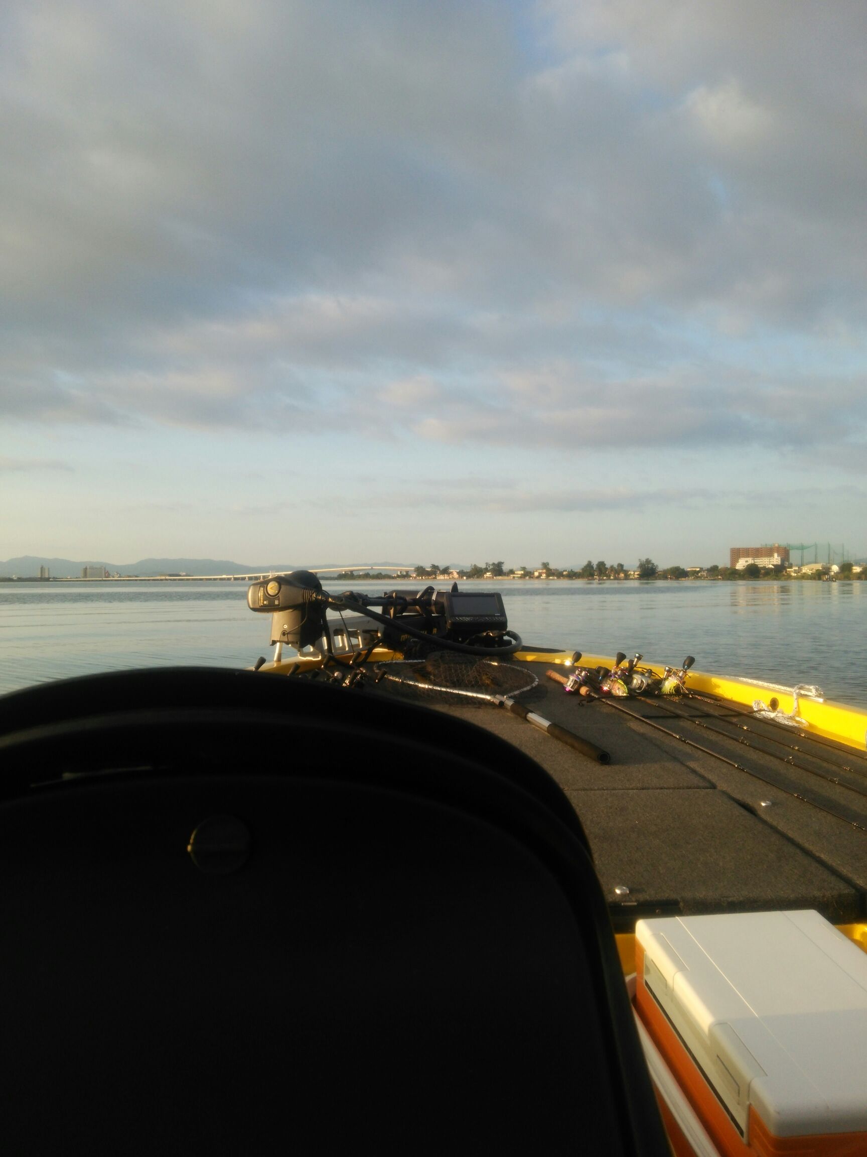
行って来ました。京都府南部にある、ボートでトラウトが釣れる管理釣り場『通天湖』へ。
この時期、いつも大放流をされるのでホームページをチェックしてみると金曜日が放流、で自分の休みが土曜日!
これは行きたい!しかし、土曜日は子供に左右されるのが常々。とりあえず、お姉チャンに予定を聞いてみた。
「釣り行きたい。」
なんと、親父の思いを知ってか知らずか最高の返答が!ありがとう、ありがとう、どうぶつの森。
ということで向かった通天湖。道中は前日に降った雪で積雪もあり、釣り場も雪景色。
昼前からスタート。とりあえずキャストを教えるところから始まり、重めのスプーンで広く探りますがマスさんは口を使ってくれません。
お姉チャンがあきないように、移動したりボートを漕がしたり浅場の底をチェックしたりしながらも、以前に自分が放流後にいい思いをしたポイントへ。
これが大正解。1投目からフェザージグにレインボーが、2投目クランクにも。
さらに1.6gスプーンにも釣れてきて、どうも中層で浮いている感じ。
お姉チャンもテンション上がって投げるも、木に引っかかったりで、なかなか掛からず。
しかし、ホスト役に徹してコチラが巻いて止めてを教えると早々にヒット!
その後も掛かる→ばらすを何回か繰り返し、充分楽しんで時間となりました。
結果、お姉チャンも釣れて自分も満足した釣果に良い釣りができました。
「良かったなぁ釣れて。また付いて行ってあげるわ」
と帰りの車で、お褒めの言葉を頂きました。


