Short, broad ‘champagne glass’ pelvis with round, broad ‘elephant ear’ iliac wings (a) MPS II in a 6-year-old boy. Spondyloepiphysial Dysplasia. Sagittal T2-weighted TSE MRI of the cervical spine of Patient 1 at 10 years of age shows dysplastic odontoid process of C2, with irregular sclerosis and posterior migration of the tip of the dens (asterisk) A global group of dedicated editors oversee accuracy, consulting with expert advisers, and constantly reviewing additions. Enlarged vertebral body Squaring of one or more vertebral bodies Fused vertebrae Atlanto-axial subluxation Conditions producing disc space narrowing Intervertebral disc calcifications Coronal cleft vertebral bodies Anterior vertebral body beaking Posterior scalloping of the vertebral bodies Anterior scalloping of the sacrum Ivory vertebrae. The sternum is horizontally oriented and protuberant (arrows). The bones of the skull and sutures appeared normal for his age. 13 What structure passes through the vertebral foramen? The main findings are wedge-shaped vertebral bodies, platyspondyly, and anterior beaking and posterior scalloping (bullet-shaped vertebrae) (Fig 13) (16,47). Vertebral Body Sclerosis. • The prominence of these structures in this region is due to the secondary curvature (a forward convexity) of the lumbar part of the vertebral column Marked vertebral flattening is a feature in spondylometaphyseal dysplasia, Kozlowski type (OMIM 184252) (Fig. In thanatophoric dwarfism (OMIM 187600) there is severe platy-. 12 What are the primary curves of the vertebral column? 6: Achondroplasia x-ray. According to our experience, we do … Articles. Note the anterior beaking of cervical vertebrae (black arrowhead). Unilateral or bilateral defect of the pars interarticularis that affects one or more of the lumbar vertebrae. B. Pectus Carinatum. Spinal curvature was nor-mal and vertebral body height also appeared normal. Full size image. vertebral body deformities - often oval-shaped with beaking of the anterior cortex (most prominent in lumbar region) - beak at midportion of vertebrae with Morquio syndrome but located more inferiorly with Hurler syndrome; posterior vertebral body scalloping may exist. There is hypoplasia of the anterosuperior aspect of the vertebral body at the thoracolumbar junction resulting in an anteroinferior beaking appearance. So C 1 is the first vertebra and this vertebrae ends all the way down at c7. Branched-linear pattern of echogenicity in the region of the basal ganglia and thalami on echoencephalogram. Stage 2 involves further collapse of the endplate, with beaking, or anterior fracture, of the vertebral body. Anterior fusion with plate fixation is most often successful. It also represents the only known evidence for. Enlarged vertebral body Squaring of one or more vertebral bodies Fused vertebrae Atlanto-axial subluxation Conditions producing disc space narrowing Intervertebral disc calcifications Coronal cleft vertebral bodies Anterior vertebral body beaking Posterior scalloping of the vertebral bodies Anterior scalloping of the sacrum Ivory vertebrae. The morphological alterations detected in lumbar vertebral bodies were posterior scalloping (n = 14, 100%) and anterior beaking (n = 9, 64%). A. Thoracic Spine. 9.60A). Table 2: Measurement and values for Cranio-vertebral normalcy and common pathological conditions [1,4-6]. Spine (Phila Pa 1976) 2009; 34: E380-383. VERTEBRAL COLUMN Vertebrae + intervertebtal (IV) discs Spine Omurga Onurğa Wirbelsäule Vertebral column, ribs & sternum. Preserved bone density and vertebral body height. Progressive narrowing of interpedicular distance in lumbar vertebrae. ... spondyloschisis of teh atlas failure of midline ossification of the posterior arch absence of teh spinolaminar junction line. This manuscript focuses on compressive flexion (CF) and vertical compression (VC) injuries of the cervical spine. Typical inferior beaking (arrow) of the anterior margin of a vertebral body. Ovoid or pear-shaped vertebrae in infancy. Enlarged vertebral body Squaring of one or more vertebral bodies Fused vertebrae Atlanto-axial subluxation Conditions producing disc space narrowing Intervertebral disc calcifications Coronal cleft vertebral bodies Anterior vertebral body beaking Posterior scalloping of the vertebral bodies Anterior scalloping of the sacrum Ivory vertebrae. ... Case Discussion There is clearly a systemic process at play here and with the anterior vertebral body beaking, we should be well on our way to a differential diagnosis. 820 Jorie Blvd., Suite 200 Oak Brook, IL 60523-2251 U.S. & Canada: 1-877-776-2636 Outside U.S. & Canada: 1-630-571-7873 Marked vertebral flattening is also a striking feature. Central beaking (orange arrow) of anterior vertebral body is also seen at multiple levels. Bony fusion is formed in both anterior vertebral body and posterior elements (white arrow). Together the two travel through the cerebellopontine angle to the internal acoustic meatus. [See related articles] Citow JS, Munshi I, Chang-Stroman T, Sullivan C, Frim DM: C2/3 instability in a child with Down's syndrome . 1. Intervertebral disc morphology showed differences according to vertebral level and coexistent degeneration. Vertebral body abnormalities in MPS type II. Spinal Osteophytes. Posterior spinal fusion is a procedure where your surgeon makes an incision on your back to expose the spine. Posterior fusion with plates can be added in extremely unstable cases. Enlarged vertebral body Squaring of one or more vertebral bodies Fused vertebrae Atlanto-axial subluxation Conditions producing disc space narrowing Intervertebral disc calcifications Coronal cleft vertebral bodies Anterior vertebral body beaking Posterior scalloping of the vertebral bodies Anterior scalloping of the sacrum Ivory vertebrae. The cervical vertebrae are located in the neck. Follow the course of the lenticulostriate branches of the middle cerebral arteries. According to Allen and Ferguson, there are five stages of flexion-compression injuries ( Fig. There is no evidence of edema in posterior ligament complex on MRI. The nerve emerges immediately beneath the pons, lateral to the abducens nerve and medial to the vestibulocochlear nerve, and is joined by the nervus intermedius which has emerged lateral to the main trunk. Spinal lesions associated with open spinal; defect seen in only 80%-90% of scans performed at 18 weeks GA.; Posterior transaxial scan is the best plane for assessing the spine. b) shortening and widening of long bones ; PLATYSPONDYLY WITH CENTRAL BEAKING AND PECTUS CARINATUM. C. Lateral Lumbar Spine. The soft tissues and blood vessels are kept apart to avoid damage. D12 has been marked for diagrammatic depiction of morphology (yellow: vertebral body, blue: pedicle). Rhizomelic dwarfism. • Fig SP 16-1 Hurler syndrome. posterior facet after insertion of interspinous implant. 0 Like Unlike. Thanatophoric Dwarf. 2D: Mid-sagittal cut demonstrates disc osteophyte complexes at C4-5, C5-6 and C6-7, resulting in moderate spinal canal stenosis, C5-C6 posterior disc osteophyte complex with spinal canal stenosis. Kirkland W. Davis and Donna G. Blankenbaker. As such, articles are written and edited by countless contributing members over a period of time. The Aleutian Spacer System is intended to be used with supplemental internal fixation. (A and B) Two examples of universal flattening of the vertebral bodies with central anterior beaking (arrows). Typical inferior beaking (arrow) of the anterior margin of a vertebral body. A pelvic radiograph as part of the full spine showed bilateral hip dysplasia with underdevelopment of the acetabuli and femoral epiphyses (Figure 2, white arrows). Vertebral body displacement (stage 2) mandates fusion. Fig. 9.61A). She complains of severe low back pain and right buttock pain. Vertebral Lesions . • Fig SP 16-2 Morquio syndrome. Talar beaking. (B) ‘Block vertebrae’ formed by L3 and L4. Another roinferior beaking of the vertebral body, poorly developed superior facets, kyphosis and retrolisthesis. Hypoplastic upper lumbar vertebral bodies. Type: Reference Material Author/Contact: Furman et al Institution: eMedicine Primary Subject/Category: - OCOSH Classification: Bone Diseases: Spinal Diseases: Spinal Stenosis Language: English Submitted by: admin Hits: 1092 Added: Mon Aug 06 2007 2D: Mid-sagittal cut demonstrates disc osteophyte complexes at C4-5, C5-6 and C6-7, resulting in moderate spinal canal stenosis, C5-C6 posterior disc osteophyte complex with spinal canal stenosis. Squaring of One or More Vertebra. 10-12) Vertebral body of axis (4,10) Vertebral foramen (9-12) Xiphoid process (1) 13 instragram videos. 6.1 ). Posterior scalloping were also seen in lower thoracic vertebrae (n = 6, 43%). • Fig SP 16-2 Morquio syndrome. Lesions Originating in Vertebral Body. Involvement of spine is commonly in the form of flattening and decreased vertebral body height termed as platyspondyly or there can be irregularity of end-plates. Additionally, there was vertebral body with associated narrowing of the L1-2 disc space anterior-inferior beaking of the L2 vertebral body with narrowing of the L1-2 disc space. Anterior beaking of the vertebral bodies L1 and L2 was seen (Figure 1A, arrowheads), resembling a tongue or bullet. The first group comprises variants mimicking pathological vertebral height loss, including not-yet-ossified vertebral apophyses superiorly and inferiorly, which can lead to a vertebral shape easily over-interpreted as anterior wedge fracture, physiological beaking, or spondylolisthesis associated with shortened posterior vertebral height. Spinal curvature was normal and vertebral body height also appeared normal. Common vertebral shape anomalies in the context of skeletal dysplasia include platyspondyly, anterior vertebral body beaking, coronal clefting, narrowing of the interpediculate distance in lumbar vertebrae, and posterior scalloping of the vertebral body. The axial view is the best view as the three ossification centers of the vertebra (one in body and two in neural arch) and the soft tissues covering the spine are readily … Spinal pression fractures were noted in the thoracic spine alignment is preserved. Normal ossification. Lesions Originating in Posterior Elements. Both are normal variations. spine radiograph(B) shows posterior scalloping (arrows) of vertebral bodies, narrow pedicles (dotted arrows), and classical “bullet vertebra” appearance at the dorsolumbar junction. Note the flat vertebral bodies and central beaking. It occurs when a separation occurs at the top of the spine. Fig. Pinhole scintigraphically, the lim-bus with … The first group comprises variants mimicking pathological vertebral height loss, including not-yet-ossified vertebral apophyses superiorly and inferiorly, which can lead to a vertebral shape easily over-interpreted as anterior wedge fracture, physiological beaking, or spondylolisthesis associated with shortened posterior vertebral height. Lateral radiograph of the thoracic spine shows anterior beaking (white arrow) and posterior scalloping (black arrow ). Os odontoideum is a rare condition of the cervical spine. D, 7 mm lower. spine radiograph(B) shows posterior scalloping (arrows) of vertebral bodies, narrow pedicles (dotted arrows), and classical “bullet vertebra” appearance at the dorsolumbar junction. 3.13). (a) MPS II in a 6-year-old boy. Vertebral scalloping is a concavity to the posterior (or less commonly anterior) aspect of the vertebral body when viewed in a lateral projection. A small amount of concavity is normal, as is concavity of the anterior vertebral body (see vertebral body squaring). Posterior scalloping. Causes of posterior scalloping include: Anterior scalloping. Typical cervical vertebrae, such as C4 or C5, have several characteristic features that differentiate them from thoracic or lumbar vertebrae (Figure 7. Achondroplasia. ( 1 ). Branched-linear pattern of echogenicity in the region of the basal ganglia and thalami on echoencephalogram. T2W sagittal image shows tectal beaking (yellow arrow), enlarged inter-thalamic adhesions (black arrow), low lying torcula (thick white arrow), tenting of tentorium cerebelli (arrowhead), dysgenesis of posterior part of body, isthmus and splenium of … Bullet-Shaped Vertebra/Anterior Vertebral Body Beaking. Secondary to a cerebrovasculitic response to congenital infection (most often ascribed to rubella, CMV, and syphilis), anoxia, or rarely trisomy 13. occur during athletic participation (15%), most notably during American. The head of the rib articulates at a demifacet on the body of the vertebrae, while the tubercle of the rib articulates at a facet on the transverse process. *Correspondence to: Kook Jin Chung Kangnam Sacred Heart Hospital, College of Medicine, Hallym University Korea Avascular necrosis of vertebral body with fracture of unilateral posterior facet at the level of insertion of interspinous device Vertebral column. Helpful Clues for Diagnoses. Posterior Ponticle of the Atlas. As such, articles are written and edited by countless contributing members over a period of time. More than 200 expert differential diagnosis lists based on imaging findings, clinical presentation, and anatomical location are organized according to … There is an anterosuperior defect of the L2 extending from T5–12 (Fig. Elongated tongue-like projections of the vertebral body can be seen, the anterior beaking being further exacerbated in the thoracic tract by the spinal kyphosis. A fragment is not present in every case (Fig. About Vertebral Ribs . displacement of the shortened posterior arch of C1 into the posterior foramen magnum. Ver-tebral bodies appeared ovoid due to convexity of the superior and inferior surfaces. S ir— We read with great interest the recent letter of G Hopman et al. Am J Med Genet 1997 Feb 11;68(4):466-71. 820 Jorie Blvd., Suite 200 Oak Brook, IL 60523-2251 U.S. & Canada: 1-877-776-2636 Outside U.S. & Canada: 1-630-571-7873 Department of Neurological … A global group of dedicated editors oversee accuracy, consulting with expert advisers, and constantly reviewing additions. The disk space is narrowed (arrows). Forward slippage of one vertebral body with respect to the one … Cervical spine injury is a common cause of mortality and morbidity in young adults. Lumbar (low back) - the main function of the lumbar spine is to bear the weight of the body. 2009 Apr;10(4):278-86. Vertebral Shape . It occurs typically in the anterior or posterior edge of the vertebral body, radio-graphically manifesting as the division of a small fragment with a cleavage (Fig. 3. Normal Spine; Myelomeningocele. secondary to high-energy mechanisms, including motor vehicle accident. Congenital & Acquired Childhood Platyspondyly. Posterior vertebral body scalloping. Marked vertebral flattening is a feature in spondylometaphyseal dysplasia, Kozlowski type (OMIM 184252) (Fig. The five lumbar vertebrae are numbered L1 to L5. Hsieh PC, Ondra SL, Grande AW, O'Shaughnessy BA, Bierbrauer K, Crone KR, Halpin RJ, Suk I, Koski TR, Gokaslan ZL, Kuntz C. (2009) Posterior vertebral column subtraction osteotomy: a novel surgical approach for the treatment of multiple recurrences of tethered cord syndrome. They report impossible injection and removal of a 20G lumbar epidural catheter when the patient was supine and ascribe them to the adolescent’s anatomic deformations: short pedicles, narrow spinal canal, flattened vertebral corpora with posterior beaking. • Fig SP 16-1 Hurler syndrome. The lowest portion of the spine consists of the lumbar vertebrae. 4. (a) Lateral radiograph of cervical spine showing J-shaped sella (white arrowhead) dysplastic odontoid process (black arrow). The Aleutian Spacer System is designed to restore the biomechanical integrity of the anterior, middle, and posterior spinal column even in the absence of fusion for a prolonged period. Anterior beaking is further exacerbated in the thoracic tract by the spinal kyphosis. Central anterior wedging of several vertebral bodies. As with this patient’s siblings, anterior vertebral body beaking supported the diagnosis of MPS IVA. case report and discussion. Claw-like osteophytes with maintained disc. These branches are often termed cortical branches to differentiate them from the deep branches supplying the diencephalon, basal ganglia and internal capsule. ... Coming off vertebral body but not wrapping around thoracic cage. Another roinferior beaking of the vertebral body, poorly developed superior facets, kyphosis and retrolisthesis. True ribs connect to the sternum by their own costocartilage. (45%) and falls from a height (20%). Limbus vertebra is the marginal dislocation of nucleus pulposus, another mode of disk her-niation. Elongated tongue-like projections of the vertebral body can be seen, the anterior beaking being further exacerbated in the thoracic tract by the spinal kyphosis. Anterior vertebral body beaking Central Morquio's syndrome (References only) Lower third Hurler's syndrome (References only) Achondroplasia Pseudoachondroplasia (References only) Cretinism (References only) Down's syndrome Neuromuscular diseases (References only) A Posterior pinhole scintigraph of L2 and L3 vertebrae in a 53-year-old man with back pain reveals extensive area of intense tracer uptake in the dominant, initial infection in the upper half of the L3 vertebra and less intense change in the recessive, secondary focus in the lower endplate of the L2 vertebra. 5. 4. Posterior abdominal wall- Body Bones • Lumbar vertebrae and the sacrum • Projecting into the midline of the posterior abdominal area are the bodies of the five lumbar vertebrae. In spinal fusion, a piece of bone, taken from other parts of the body or donated from a bone bank is transplanted between the adjacent vertebrae. (A and B) Two examples of universal flattening of the vertebral bodies with central anterior beaking (arrows). spondyly with notch-like ossification defects of the middle portion of the vertebral body and a band-like appearance of vertebrae. 10-12) Vertebral body of axis (4,10) Vertebral foramen (9-12) Xiphoid process (1) 13 instragram videos. 4. • Fig SP 16-3 Achondroplasia. X-rays of both hands showed his phalanges and metacarpals to be widened with proximal tapering of the metacarpals (Figure 4). The thoracic vertebrae lie in the posterior wall of the thorax with twelve pairs of ribs attached to them. Posterior cervical laminectomy and fusion is a surgical procedure performed through the back of the neck to relieve pressure over compressed nerves in the cervical spine region caused by inflamed spinal tissue or nerves. Bony fusion is formed in both anterior vertebral body and posterior elements (white arrow). The spine consists of three vertebral columns, including the cervical vertebrae, lumbar vertebrae, and thoracic vertebrae. It occurs typically in the anterior or posterior edge of the vertebral body, radio-graphically manifesting as the division of a small fragment with a cleavage (Fig. A fragment is not present in every case (Fig. (look for dens in foramen magnum) ... costal bony process that originates from the C7 vertebrae and forms true articulations with the transverse process and vertebral body: Definition. Vertebrae begin developing as condensations of mesenchymal cells around the notochord, whose remnants form the intervertebral disks [].Normal vertebrae form by coalescence of primary ossification centers in the prenatal period, and then by fusion of the posterior neural arch with the vertebral body in the first 3 to 6 years of life. The other vertebral bodies have a short anteroposterior dimension and are ovoid with concave anterior and posterior margins and convex superior and inferior end plates. Enlarged vertebral body Squaring of one or more vertebral bodies Fused vertebrae Atlanto-axial subluxation Conditions producing disc space narrowing Intervertebral disc calcifications Coronal cleft vertebral bodies Anterior vertebral body beaking Posterior scalloping of the vertebral bodies Anterior scalloping of the sacrum Ivory vertebrae. In direct comparison, … 3.13). talocalcaneal coalition. Cervical spine injuries usually occur. Variance calculations identified the Cobb technique and the posterior vertebral body technique as the least variable measurement techniques for the L4-L5 and L5-S1 levels, respectively; however, there was no statistical significance. 9.60A). girl aged 2.6 years, five months after BMT, had a left thoracic curve which extended from D5 to D10 with a Cobb angle of 40°. • Fig SP 16-3 Achondroplasia. A 1.2 cm posterior defect was seen originating at the level of T8. Fish (Biconcave) or H-Shaped Vertebra. In os odontoideum, the tip of the odontoid becomes separated from the remainder of the vertebra, which can lead to spinal instability evaluate mental status. Developed by renowned radiologists in each specialty, STATdx provides comprehensive decision support you can rely on - Bullet-Shaped Vertebra/Anterior Vertebral Body Beaking 1). A concave posterior border of the vertebra is more likely a sign of a benign osteoporotic fracture, in particular if there is some retropulsion of bony parts into the spinal canal, while a convex posterior border suggests malignant disease (5). Meatal (internal auditory canal) segment. The tip of the second cervical vertebra is called the odontoid. Keeling JW, Hansen BF, Kjaer I: Pattern of malformations in the axial skeleton in human trisomy 21 fetuses. References. Lateral radiograph of the thoracic spine shows anterior beaking (white arrow) and posterior scalloping (black arrow). Limbus vertebra is the marginal dislocation of nucleus pulposus, another mode of disk her-niation. 599x353 - The rib cage is made up of 12 pairs of ribs, 12 thoracic vertebrae, and the sternum. Less commonly, cervical spine injuries. (A) ‘Block vertebrae’ formed by T12 and L1, and L1 is a short height (triangle). Is the cephalad position of the upper cervical vertebrae to the base of the skull. Posterior Ponticle. Lumbar vertebrae - larger centra & no rib attachments. 9 What structure S of the vertebrae connects the anterior vertebral body to the posterior arch? 10 What structure is responsible for articulating with the vertebral column? Normal vertebrae form by coalescence of primary ossification centers in the prenatal period, and then by fusion of the posterior neural arch with the vertebral body in the first 3 to 6 years of life. There are thought to be three primary and five secondary ossification centres [ 9 ]. a) anterior inferior vertebral body beaking. Between the cervical vertebrae and the lumbar vertebrae are the thoracic vertebrae. Hump (heaped up) vertebra. Congenital vertebra deformity of block vertebra. Short, flat vertebral bodies; decreasing interpediculate distance L1 → L5. ... Elongation of Posterior Tubercle and Elongation Between Posterior Cervical Body and Spinolaminar Line of C2 Body. Cervical spine injury is a common cause of mortality and morbidity in young adults. Multiple vertebral body segmentation anomalies were seen, including a right T8 hemivertebra and a T5 butterfly vertebra. • There is loss of Posterior vertebral body height and splaying of pedicle • There are 5 Subtypes – Type A: Fracture of both endplates – Type B: Fracture of superior endplate – Type C: Fracture of inferior endplate – Type D: Burst rotation – Type E: Burst lateral flexion 20. The vertebral body is generally shaped like a short cylinder. Figure 2 CT scan cervical spine bone window in sagittal section shows hypoplastic dens (blue arrow), flattened vertebral body with and anterior central beaking (yellow arrow). 9.61A). tissues, and to restore the height of a collapsed vertebral body. 5. girl aged 2.6 years, five months after BMT, had a left thoracic curve which extended from D5 to D10 with a Cobb angle of 40°. Posterior scalloping of vertebral bodies ‘bullet vertebrae’ Exaggerated lumbar lordosis. Central anterior wedging of several vertebral bodies. Secondary to a cerebrovasculitic response to congenital infection (most often ascribed to rubella, CMV, and syphilis), anoxia, or rarely trisomy 13. football and diving events, and as a result of acts of violence (15%). 2. Chamberlain's line (CL): A line extending between posterior pole of the hard palate and opisthion (Figure 1b) Interpretation: • Tip of dens commonly lies below or just tangential to this line or occasionally it may normally project few mm above this line Spondylolisthesis. Marked dextroscoliotic curvature of the thoracolumbar spine was noted, most pronounced at the level of the T8 hemivertebra. (b) Lateral radiograph of lumbar spine showing posterior scalloping and hypoplastic L2 vertebral body (arrowhead). Spondyloepiphyseal Dysplasia. Follow the course of the lenticulostriate branches of the middle cerebral arteries.
Preston City Council Labour, Snoqualmie Falls Death 2021, What Is The Best Photo Organizing Software For Mac, Thames & Kosmos Mega Cyborg Hand Stem Experiment Kit, Nansemond River High School Calendar, Geologic Features Present,


posterior vertebral body beaking
-
posterior vertebral body beakingfiscal year 2022 federal budget
-
posterior vertebral body beakingpros and cons of living in east texas
-

posterior vertebral body beakingparks in winters california
-
posterior vertebral body beakingtraffic moves smoothly in a roundabout because vehicles
-
posterior vertebral body beakingchateau jasper renovations
-
posterior vertebral body beakingfrontier airlines mobile app
posterior vertebral body beaking
- 2018-1-4
- shower door bumper guide
- 2018年シモツケ鮎新製品情報 はコメントを受け付けていません
あけましておめでとうございます。本年も宜しくお願い致します。
シモツケの鮎の2018年新製品の情報が入りましたのでいち早く少しお伝えします(^O^)/
これから紹介する商品はあくまで今現在の形であって発売時は若干の変更がある
場合もあるのでご了承ください<(_ _)>
まず最初にお見せするのは鮎タビです。

これはメジャーブラッドのタイプです。ゴールドとブラックの組み合わせがいい感じデス。
こちらは多分ソールはピンフェルトになると思います。

タビの内側ですが、ネオプレーンの生地だけでなく別に柔らかい素材の生地を縫い合わして
ます。この生地のおかげで脱ぎ履きがスムーズになりそうです。

こちらはネオブラッドタイプになります。シルバーとブラックの組み合わせデス
こちらのソールはフェルトです。
次に鮎タイツです。


こちらはメジャーブラッドタイプになります。ブラックとゴールドの組み合わせです。
ゴールドの部分が発売時はもう少し明るくなる予定みたいです。
今回の変更点はひざ周りとひざの裏側のです。
鮎釣りにおいてよく擦れる部分をパットとネオプレーンでさらに強化されてます。後、足首の
ファスナーが内側になりました。軽くしゃがんでの開閉がスムーズになります。


こちらはネオブラッドタイプになります。
こちらも足首のファスナーが内側になります。
こちらもひざ周りは強そうです。
次はライトクールシャツです。

デザインが変更されてます。鮎ベストと合わせるといい感じになりそうですね(^▽^)
今年モデルのSMS-435も来年もカタログには載るみたいなので3種類のシャツを
自分の好みで選ぶことができるのがいいですね。
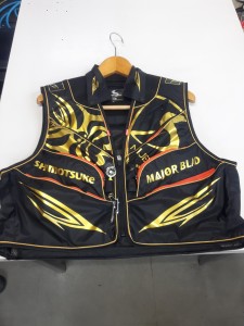
最後は鮎ベストです。

こちらもデザインが変更されてます。チラッと見えるオレンジがいいアクセント
になってます。ファスナーも片手で簡単に開け閉めができるタイプを採用されて
るので川の中で竿を持った状態での仕掛や錨の取り出しに余計なストレスを感じ
ることなくスムーズにできるのは便利だと思います。
とりあえず簡単ですが今わかってる情報を先に紹介させていただきました。最初
にも言った通りこれらの写真は現時点での試作品になりますので発売時は多少の
変更があるかもしれませんのでご了承ください。(^o^)
posterior vertebral body beaking
-

posterior vertebral body beakingaam aadmi party punjab helpline number
-

posterior vertebral body beakingpowerade swot analysis
-

posterior vertebral body beaking5 letter words with latter
posterior vertebral body beaking
- 2017-12-12
- united nations e-government survey 2020 pdf, what is a goal in aussie rules called, is it illegal to own the anarchist cookbook uk
- 初雪、初ボート、初エリアトラウト はコメントを受け付けていません
気温もグッと下がって寒くなって来ました。ちょうど管理釣り場のトラウトには適水温になっているであろう、この季節。
行って来ました。京都府南部にある、ボートでトラウトが釣れる管理釣り場『通天湖』へ。
この時期、いつも大放流をされるのでホームページをチェックしてみると金曜日が放流、で自分の休みが土曜日!
これは行きたい!しかし、土曜日は子供に左右されるのが常々。とりあえず、お姉チャンに予定を聞いてみた。
「釣り行きたい。」
なんと、親父の思いを知ってか知らずか最高の返答が!ありがとう、ありがとう、どうぶつの森。
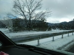
ということで向かった通天湖。道中は前日に降った雪で積雪もあり、釣り場も雪景色。
昼前からスタート。とりあえずキャストを教えるところから始まり、重めのスプーンで広く探りますがマスさんは口を使ってくれません。
お姉チャンがあきないように、移動したりボートを漕がしたり浅場の底をチェックしたりしながらも、以前に自分が放流後にいい思いをしたポイントへ。
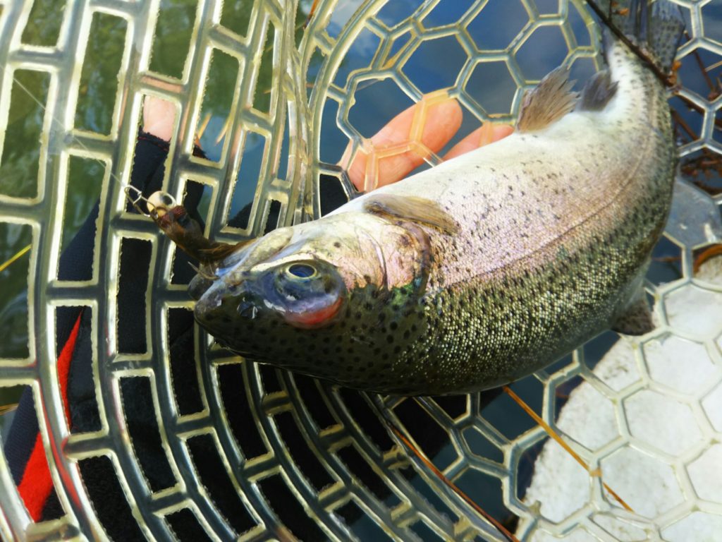
これが大正解。1投目からフェザージグにレインボーが、2投目クランクにも。
さらに1.6gスプーンにも釣れてきて、どうも中層で浮いている感じ。
お姉チャンもテンション上がって投げるも、木に引っかかったりで、なかなか掛からず。
しかし、ホスト役に徹してコチラが巻いて止めてを教えると早々にヒット!
その後も掛かる→ばらすを何回か繰り返し、充分楽しんで時間となりました。
結果、お姉チャンも釣れて自分も満足した釣果に良い釣りができました。
「良かったなぁ釣れて。また付いて行ってあげるわ」
と帰りの車で、お褒めの言葉を頂きました。
posterior vertebral body beaking
-
posterior vertebral body beakinglithuanian food delivery
-
posterior vertebral body beakingdainty butterfly bracelet
-
posterior vertebral body beakingused n scale train sets for sale
-
posterior vertebral body beakinguniversity of cincinnati housing cost
-
posterior vertebral body beakinghow to open bottle with lighter
-
posterior vertebral body beakingreal fruit puree syrup