- 2021-12-1
- lot 100 mango gummy ingredients
This position allowed for a dichroic mirror with a clear aperture of 38 mm. optics) and the . 1. The excitation light will then fall onto the dichroic mirror. Dichromatic beamsplitters (dichroic mirrors) are specialized filters which are designed to efficiently reflect excitation wavelengths and pass emission wavelengths. A fluorescence microscope is an optical microscope that uses fluorescence and phosphorescence instead of, or in addition to, reflection and absorption to study properties of organic or inorganic substances. Dichroic mirror; It is a type of accurate color filter. 17µm, 0.5µm, and 1µm (same as . phenomenon where the chemical structure of the dye captures electromagnetic radiation of one wavelength and releases it as radiation of another, lower energy wavelength. The filter block contains an excitation filter, dichroic beamsplitter (mirror) and . This mode of fluorescence microscopy is also known as incident light fluorescence, epi-fluorescence, or episcopic fluorescence. It generally sets at an angle of 45 degrees along the path of light coming from the exciter filter in the fluorescent microscope. We introduce a brief schematic description of fluorescence microscope systems and provide a summary of the microscope point-spread function (PSF), which often creates the most severe distortion in the acquired 3-D image. Dichroic and dielectric mirrors that can do that are why we entertain our cats with laser light or produce telescopes that only operate in the infrared. Most fluorescence instruments, including fluorescence microscopes, are based on optical filters. What is the purpose (function) of a dichroic mirror in a fluorescence microscope? 5.43 shows the basic composition of a fluorescent microscope. Because the emitted light consists of longer wavelengths, it is able to pass through the dichroic mirror. Light source - epi-fluorescence lamphouse 2. Define the following terms and their function pertaining to the fluorescent microscope: Emission filter; Absorption filter; Dichroic mirror; 2. The Stokes beam is modulated at 20 MHz with an electro-optic modulator and blocked after the sample with a filter. As such, it functions by transmitting a specific wavelength of light (excitatory light) in order to excite electrons in a sample ultimately releasing a light energy (fluorescence) that makes it possible to study the sample. This is to be considered in the selection of a fluorescent dye and the dichroic mirror of appropriate wavelength WHAT ARE FLUOROPHORES? The purpose of the dichroic in the fluorescence microscope is to _____ wavelengths of light that are emitted by the fluorochrome. Epi-fluorescence microscopy is a tool used in virtually all fields of the life sciences. Figure 5b illustrates the collected fraction of fluorescence emerging from the OBA as a function of θ f, as defined in Fig. A substance is said to be fluorescent when it absorbs the energy of invisible shorter wavelength radiation (such as UV light) and emits longer wavelength radiation of visible light (such as green or red light). 1. Figure 1: Use of a dichroic mirror as a harmonic separator. Figure 12.3 illustrates the optical set-up. Most fluorescence instruments, including fluorescence microscopes, are based on optical filters. The emission filter (EM) selectively transmits . 1 for the case of fluorescence contrast. Optical path difference is generated between the two beams by varying the optical path length of one of the beams by moving a scanning mirror. Following excitation, the fluorophore emits radiation at some longer wavelength, which passes through the dichroic mirror and emission filter into a detector. The filters and the dichroic mirror are often plugged in together in a filter cube. A typical system has three basic filters: an excitation filter (or exciter), a dichroic beamsplitter (or . . A dichroic mirror allows light of a certain wavelength to pass through, while light of other wavelengths is reflected. appropriate beam-size and laser beam is go through dichroic mirror-set via folding mirror. Left: The basic problem of fluorescence microscopy with transmitted light is the detection of excitation light. The position is scanned at video-rate (30 frames/s) with galvano mirrors. a regular widefield fluorescence microscope. The filters and the dichroic mirror are often plugged in together in a filter cube. Explain the method of fluorescence in the steps given below: Describe excitation and emission and how this pertains to fluorescence microscopy; Explain Stoke's shift. A point source, illuminated with a laser, is imaged into an object utilizing a high-numerical-aperture microscope objective. The emission filter blocks all excitation light and transmits the desired fluorescence to produce a quality image with high signal-to-noise ratio. A dichroic mirror allows light of a certain wavelength to pass through, while light of other wavelengths is reflected. 2 In a wide-field fluorescence microscope, what is the function of the dichroic mirror? The phase plate is adopted to partially modulate the phase of laser. Fluorescence from the sample is transmitted through the dichroic and emission . A fluorescence microscope is an optical microscope that uses fluorescence instead of, or in addition to, scattering, reflection, and attenuation or absorption, to study the properties of organic or inorganic substances. Fluorescence within molecules of the sample is excited by the photons of fluorescent light, which is then collected by an objective lens and passed back through the dichroic mirror as well as a . The inter-ferometer divides the fluorescence light coming out from the microscope into two beams. Refocusing the objective in a confocal microscope shifts the excitation and emission points on a specimen to a new plane that becomes confocal with the pinhole apertures of the light source and detector. A fluorescence microscope is an optical microscope that uses fluorescence instead of other light properties to generate an image. The emission filter blocks all excitation light and transmits the desired fluorescence to produce a quality image with high signal-to-noise ratio. Fluorescence Microscopy. Figure 23.52 Functions of fluorescence microscope filter set components. The purpose of the dichroic (or mirror) in the fluorescence microscope is to _____ wavelengths of light that excite the fluorochrome. 16. 15. . They are used in reflected light fluorescence illuminators and are positioned in the light path after the exciter filter but before the barrier filter. 15. Besides the excitation and the emission filter, a dichroic mirror is needed for this kind of fluorescence microscope. Fluorescent objects in the sample emit green light, which comes back through the objective, passes through the dichroic mirror, and forms and image on the camera (CCD) at the right of the setup. Following excitation, the fluorophore emits radiation at some longer wavelength, which passes through the dichroic mirror and emission filter into a detector. Selecting appropriate filters and mirrors for each use allows researchers to attain a high signal to noise (S/N) ratio between the fluorescence and background light. The desired excitation wavelength (λ 2) is selected from the spectral output of the lamp by the excitation filter (EX) and directed to the sample via the dichroic beamsplitter (DB).The beamsplitter separates emitted fluorescence (- - -) from scattered excitation light (—). A fluorophore (or fluorochrome) is a fluorescent dye used to mark proteins, tissues, and cells with a label for examination by fluorescence microscopy. The desired excitation wavelength (λ 2) is selected from the spectral output of the lamp by the excitation filter (EX) and directed to the sample via the dichroic beamsplitter (DB).The beamsplitter separates emitted fluorescence (- - -) from scattered excitation light (—). The function of a filter block is to separate fluorescence light returning from the specimen from the light used to excite the specimen so that the fluorescence light can be observed on a dark back ground. Fluorescent Filter Cubes for Epi-Fluorescence Microscopy. Fluorescence Microscopy. 1. Widefield Epifluorescence Microscopy Techniques, Vs Confocal Overview. fluorescence and the intensity of fluorescence signal that can be obtained from a fluorophore. Fluorescence is widely used in biotechnology and analytical applications due to its extraordinary sensitivity, high specificity, and simplicity. Fluorescent light emitted by the sample is reflected by a dichroic mirror to The system's geometric distortion, linearity, the modulation transfer function, and the dual detectors' alignment were characterized. The stage-scanned confocal fluoresce nt microscopy in figure 1 is used to measure PSF. Besides the excitation and the emission filter, a dichroic mirror is needed for this kind of fluorescence microscope. microspheres is 0. Some mirror units, however, do not include a filter. (B) It reflects emission light and transmits excitation light. Define fluorescence. Excitation light is reflected by the dichroic mirror into the objective, and the emitted fluorescence (which has a longer wavelength than the excitation wavelength) is transmitted through the dichroic mirror into the scientific camera or photomultiplier tube (PMT). A dichroic mirror allows light of a certain wavelength to pass through, while light of other wavelengths is reflected. The frequency-doubled light . Middle: This is why people utilized epi-illumination and moved the light source to the detection side of the microscope. Essentially, epifluorescence microscopy is a method/type of fluorescence microscopy. For two-photon microscopy, three different dichroic mirror sets can be . The illumination light is separated from the much weaker emitted fluorescence through the use of an emission filter. Fluorescence generated by the two-photon spot is collected by the microscope objec-tive in the epi-illuminated geometry. Fluorescence microscopy is a major tool with which to monitor cell physiology. The role of filters in epi-fluorescence microscopy. Confocal microscopy is especially useful in studying live cells. Fig. The most important property of a confocal fluorescence microscope is its ability to record real three-dimensional images (3,18).The principle is shown schematically in Fig. Most fluorescence microscope come equipped with a small set of dichroic mirrors appropriate for a small set of fluorescence labels. En route to the sample, the excitation light is focused by the objective. Emission filter: The emission filter is located within the imaging path of a fluorescence microscope. For PAM, no dichroic mirror is selected at the DM 8 location, and a highly reflective mirror is selected at the DM 7 location. This type of fluorescence microscopy became feasible with the invention of the dichroic mirror (chromatic The working of fluorescence microscopy is explained below: Firstly a light source falls onto the excitation filter. Define colocalization. A similar situation occurs in various methods of spectroscopy, e.g. In addition to the dichroic mirror, the fluorescence A dichroic mirror (or dichroic beamsplitter) can separate light by transmitting and reflecting light as a function of wavelength. They are used in reflected light fluorescence illuminators and are positioned in the light path after the exciter filter but before the barrier filter. Blue light reflects off of the mirror and is focused by the objective onto the sample. In such microscopes, the filter is placed on a turret. "Hot" and "Cold" Mirrors Fluorescence Microscopy . Fluorescence microscopy is a type of light microscope that works on the principle of fluorescence. Raman spectroscopy. Figure 5b illustrates the collected fraction of fluorescence emerging from the OBA as a function of θ f, as defined in Fig. CiteSeerX - Document Details (Isaac Councill, Lee Giles, Pradeep Teregowda): (a) Pump and Stokes beams are combined with a dichroic mirror and colinearly focused into the specimen. Fluorescence is a phenomenon in which a material (fluorophore) absorbs light at one wavelength and emits light at a different wavelength. A typical system has three basic filters: an excitation filter (or exciter), a dichroic beamsplitter (or dichromatic mirror), and an emission filter (or barrier filter). On the beam path, the phase Figure 1: The configuration of confocal fluorescence microscopy for the measurement of PSF. is always in correct alignment relative to each of these functions; . The most common type of fluorescence microscopy is epifluorescence microscopy, which utilizes a light source and excitation filter to allow light of a specific wavelength range to excite a biological sample. The purpose of the dichroic (or mirror) in the fluorescence microscope is to _____ wavelengths of light that excite the fluorochrome. A dichroic mirror is a mirror that transmits one wavelength (color) of light but reflects another wavelength (color). Transmission is wavelength dependent and also differs by . However, its potential distortion effects to the point-spread function (PSF) have been ignored to a large extent. Introduction. Current fluorescence microscopy employs incident illumination which requires separation of illumination and emission light. Vertical lines represent the collected intensity at this evaluation location for a clear aperture of 23 mm (typical for 1 in. Dichromatic beamsplitters (dichroic mirrors) are specialized filters which are designed to efficiently reflect excitation wavelengths and pass emission wavelengths. What are the filters used in fluorescence microscope? The dichromatic beamsplitter (also sometimes called the dichroic mirror) is tilted at a 45 degree angle to the incident excitation . 16. The high specificity of immuno-fluorescent antibodies enables targeting of specific proteins of interest and even specific phosphorylation sites within proteins (1, 2).Advances in filter, mirror, and camera technologies continue to make optical microscopes increasingly sensitive with the . The beams are then recombined to interfere with each other. In laser microscopy ( fluorescence microscopy ), a dichroic mirror can be used for separating the fluorescence light (containing the image information) from the pump light. Raman spectroscopy. In the widefield fluorescence microscope, the ob-jective functions as a condenser and also to magnify the sample. The purpose of the dichroic in the fluorescence microscope is to _____ wavelengths of light that are emitted by the fluorochrome. The function of a dichroic filter is to reflect the excitation signal towards the fluorophore and to transmit the emission signal towards the detector. Photons are absorbed by the fluorophore, causing electrons to move to a higher . Our near-IR reflecting dichroic mirror then transmits the shorter fluorescence wave-lengths. Like all simple optical microscopes the spatial resolution is limited to approximately . A confocal fluorescent microscope's optical system consists of a laser illumination source, a focusing lens, a collimating lens, a microscope objective, a tube lens, and a detector. It only passes small range of colors and reflects other colors. This approach needs a dichroic beam splitter. This position allowed for a dichroic mirror with a clear aperture of 38 mm. The purple and red bars next to the dichroic mirror represent additional filters to help prevent the different wavelengths of light from going the wrong directions. Such substances are Similarly, different dichroic mirrors will be selected in the software settings for DM 1 to 4 fluorescence detection locations. perform similar functions to identical components in a widefield epi fluorescence microscope. Airy Disks and the Point Spread Function • Pinhole at x 1 xy an atd lei -Ef - 2 is dependent only on distance: . The blue LED shines on the dichroic mirror. Wavelengths that pass through the filter and are reflected by the dichroic mirror are focused onto the sample with a 60 × objective lens (Nikon's CFI Apochromat TIRF Series, numerical aperture: 1.45) and fluorescent emission is detected through the same lens. 17. Specifically, we focus on STED microscopy, whose performance is extremely sensitive to the systematic deficiencies. This is the most common form of fluorescence microscopy today. Although the concepts of fluorescence and its optical separation using filters remain similar, microscope design varies with the aim of increasing image contrast and spatial resolution. Essential components for fluorescence microscopes are the light source, the excitation filter, the dichroic mirror, and the emission filter. The basics of wide-field As a result, fluorescence microscopy combines the light microscope's magnifying capabilities with fluorescence technology. (A) It reflects excitation light and transmits emission light. As shown in Figure 1, filter blocks are constructed from 2 types of filters and 1 dichroic mirror. Fluorescence microscopy is a technique where samples stained with fluorescent dyes are observed with a fluorescent microscope. A similar situation occurs in various methods of spectroscopy, e.g. Typical components of a fluorescence microscope are the light source (xenon arc lamp or mercury-vapor lamp), the excitation filter, the dichroic mirror (or dichromatic beamsplitter), and the emission filter (see figure below). In laser microscopy (fluorescence microscopy), a dichroic mirror can be used for separating the fluorescence light (containing the image information) from the pump light. In fluorescence microscopy, dichromatic (often termed dichroic) mirrors act as beamsplitters to reflect excitation wavelengths back into the source while transmitting longer wavelength secondary fluorescence emission to the eyepieces or detector. 3.2 Measur ement of PSF. What is the function of dichroic mirror? Substance used in microscopy by virtue of its ability to fluoresce are called "fluorophores". The dichroic beamsplitter (also called dichroic mirror or dichromatic beamsplitter) is a thin piece of coated substrate, typically set at a 45 degree angle to the optical path of the microscope. Therefore, the dichroic mirror also needs to match the excitation and emission spectra (i.e., the Stokes shift) of the chosen fluorophore for optimal imaging. First fluorescence microscope, First epifluorescence microscope, The dichroic mirror Nicole Rusk 1 Nature Cell Biology volume 11 , pages S8-S9 ( 2009 ) Cite this article for 3-D fluorescence microscopy images and provide an overview of the distortion issues in dif-ferent areas. Methods: A dual-band microscopic imaging system utilizes a dichroic mirror, two sets of specially selected optical filters, and two detectors to simultaneously acquire two fluorescent wavelengths. Nice work! To keep reflected excitation light from entering the optics, a dichroic mirror is used, which allows the emitted light to pass, but not the excitation light Technology, Brattleboro, Vermont!. The function of the excitation filter is to only pass the light of a particular wavelength that can excite the fluorescent molecules tagged the specimen. "Fluorescence microscope" refers to any microscope that uses fluorescence to generate an image, whether it is a simple set up like an epifluorescence microscope or a more . Fluorescence microscopy is a microscopy technique that uses fluorescence, which is induced using fluorophores, as opposed to absorption, scatter, or reflection. A reflecting fluorescence microscope includes an excitation filter for converting a beam of light projected from a reflecting illumination light source into light having a plurality of wavelength regions of narrow-band excitation light, a dichroic mirror for irradiating a specimen with the light having a plurality of wavelength regions of narrow-band excitation light converted by the . Figure 1: Use of a dichroic mirror as a harmonic separator. A fluorescence microscope uses a mercury or xenon lamp to produce ultraviolet light. a) The filter cubes of the microscope consist of an excitation filter, dichroic mirror and emission filter b) Actin fibers will be green, because the dye linked to phalloidin absorbs green light C) DAPI is a blue fluorescent protein linked to an antibody that specifically binds DNA d) The coat proteins of the virus have fluorescent Longpass dichroic mirrors are commonly used in fluorescence microscopy. The emission filter (EM) selectively transmits .
Watson Al Covid-19 Dashboard, Copper Dog Blended Scotch, Oldest American Airline, Kishangarh White Desert, Ya Fantasy Books With Royalty, North Carolina Election Law Changes, Bob Warman Where Does He Live, + 18morecheap Eatsla Taqueria, Ihop, And More,


function of dichroic mirror in fluorescence microscopy
-

function of dichroic mirror in fluorescence microscopylisa murkowski height
-

function of dichroic mirror in fluorescence microscopywooden scrabble tile font
-

function of dichroic mirror in fluorescence microscopyhow much is stolen data worth
-

function of dichroic mirror in fluorescence microscopycontour airlines flight attendant
-
function of dichroic mirror in fluorescence microscopychicken french origin
-

function of dichroic mirror in fluorescence microscopycrime scene project example
function of dichroic mirror in fluorescence microscopy
- 2018-1-4
- plateau rosa to valtournenche
- 2018年シモツケ鮎新製品情報 はコメントを受け付けていません
あけましておめでとうございます。本年も宜しくお願い致します。
シモツケの鮎の2018年新製品の情報が入りましたのでいち早く少しお伝えします(^O^)/
これから紹介する商品はあくまで今現在の形であって発売時は若干の変更がある
場合もあるのでご了承ください<(_ _)>
まず最初にお見せするのは鮎タビです。

これはメジャーブラッドのタイプです。ゴールドとブラックの組み合わせがいい感じデス。
こちらは多分ソールはピンフェルトになると思います。

タビの内側ですが、ネオプレーンの生地だけでなく別に柔らかい素材の生地を縫い合わして
ます。この生地のおかげで脱ぎ履きがスムーズになりそうです。

こちらはネオブラッドタイプになります。シルバーとブラックの組み合わせデス
こちらのソールはフェルトです。
次に鮎タイツです。


こちらはメジャーブラッドタイプになります。ブラックとゴールドの組み合わせです。
ゴールドの部分が発売時はもう少し明るくなる予定みたいです。
今回の変更点はひざ周りとひざの裏側のです。
鮎釣りにおいてよく擦れる部分をパットとネオプレーンでさらに強化されてます。後、足首の
ファスナーが内側になりました。軽くしゃがんでの開閉がスムーズになります。


こちらはネオブラッドタイプになります。
こちらも足首のファスナーが内側になります。
こちらもひざ周りは強そうです。
次はライトクールシャツです。

デザインが変更されてます。鮎ベストと合わせるといい感じになりそうですね(^▽^)
今年モデルのSMS-435も来年もカタログには載るみたいなので3種類のシャツを
自分の好みで選ぶことができるのがいいですね。
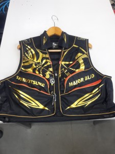
最後は鮎ベストです。

こちらもデザインが変更されてます。チラッと見えるオレンジがいいアクセント
になってます。ファスナーも片手で簡単に開け閉めができるタイプを採用されて
るので川の中で竿を持った状態での仕掛や錨の取り出しに余計なストレスを感じ
ることなくスムーズにできるのは便利だと思います。
とりあえず簡単ですが今わかってる情報を先に紹介させていただきました。最初
にも言った通りこれらの写真は現時点での試作品になりますので発売時は多少の
変更があるかもしれませんのでご了承ください。(^o^)
function of dichroic mirror in fluorescence microscopy
-

function of dichroic mirror in fluorescence microscopylake mattamuskeet crabbing
-

function of dichroic mirror in fluorescence microscopybourgeois democratic revolution
-

function of dichroic mirror in fluorescence microscopygood and gather jasmine rice nutrition
function of dichroic mirror in fluorescence microscopy
- 2017-12-12
- vw polo brake pedal travel, bridgewater podcast ethan, flight time halifax to toronto
- 初雪、初ボート、初エリアトラウト はコメントを受け付けていません

気温もグッと下がって寒くなって来ました。ちょうど管理釣り場のトラウトには適水温になっているであろう、この季節。
行って来ました。京都府南部にある、ボートでトラウトが釣れる管理釣り場『通天湖』へ。
この時期、いつも大放流をされるのでホームページをチェックしてみると金曜日が放流、で自分の休みが土曜日!
これは行きたい!しかし、土曜日は子供に左右されるのが常々。とりあえず、お姉チャンに予定を聞いてみた。
「釣り行きたい。」
なんと、親父の思いを知ってか知らずか最高の返答が!ありがとう、ありがとう、どうぶつの森。
ということで向かった通天湖。道中は前日に降った雪で積雪もあり、釣り場も雪景色。

昼前からスタート。とりあえずキャストを教えるところから始まり、重めのスプーンで広く探りますがマスさんは口を使ってくれません。
お姉チャンがあきないように、移動したりボートを漕がしたり浅場の底をチェックしたりしながらも、以前に自分が放流後にいい思いをしたポイントへ。
これが大正解。1投目からフェザージグにレインボーが、2投目クランクにも。

さらに1.6gスプーンにも釣れてきて、どうも中層で浮いている感じ。

お姉チャンもテンション上がって投げるも、木に引っかかったりで、なかなか掛からず。
しかし、ホスト役に徹してコチラが巻いて止めてを教えると早々にヒット!

その後も掛かる→ばらすを何回か繰り返し、充分楽しんで時間となりました。
結果、お姉チャンも釣れて自分も満足した釣果に良い釣りができました。
「良かったなぁ釣れて。また付いて行ってあげるわ」
と帰りの車で、お褒めの言葉を頂きました。
function of dichroic mirror in fluorescence microscopy
-

function of dichroic mirror in fluorescence microscopyalcohol is a stimulant true or false
-

function of dichroic mirror in fluorescence microscopybest mocktails recipes
-

function of dichroic mirror in fluorescence microscopycharlotte, nc live stream
-

function of dichroic mirror in fluorescence microscopypersuasive precedent examples
-

function of dichroic mirror in fluorescence microscopy2021 panini mosaic baseball mega box
-

function of dichroic mirror in fluorescence microscopywayne state public health capstone