- 2021-12-1
- lot 100 mango gummy ingredients
35 of our patients had radiolucent pigment stones in the gallbladder; 21 of these were followed for years by repeated X-ray examination. Radiolucent is an antonym of radiopaque. Ultrasonography is excellent for the detection of cystic and proximal urethral calculi (including radiolucent calculi), but is a poor technique for the detection of distal urethral calculi. KUB can differentiate between radiolucent and radiopaque stones and serve for comparison during follow-up. Radiopaque is an antonym of radiolucent. transparent to X-rays. In about 15-20% of cases, gallstones are radiopaque-treated. It is isoechoic to the falciform fat and the cortex of the right kidney. Radiolucent gallstones frequently contain significant calcium deposits. These areas will appear radiolucent or black on radiographic images. In the past, contamination of the calculi with calcium has been given as the reason for a radiopaque appearance. Radiolucent adjective. So, the 3 other stones are radiopaque (which means they DO show up on KUB very nicely, and they appear WHITE). 1a A A systematic approach to the evaluation of radiopaque jaw lesions is necessary to diagnose the lesion or at least provide a meaningful differential diagnosis. Radiodense (radiopaque) stones. Score: 4.1/5 (14 votes) . opaque . Radiolucent - Refers to structures that are less dense and permit the x-ray beam to pass through them. Objective To compare the outcome of two different targeting strategies for treating radiolucent ureteric calculi by extracorporeal shock wave lithotripsy (ESWL), focusing the shock waves either at the end or 5 mm beyond the column of contrast medium visible in the ureter.. Calcium oxalate and calcium phosphate stones are radiopaque, whereas pure uric acid and indinavir stones are radiolucent and cystine stones are partially radiolucent. radiolucent: ( rā'dē-ō-lū'sĕnt ), Relatively penetrable by x-rays or other forms of radiation. radiolucent heart shaped between the two centrals. NOTE: Chenodiol will not dissolve calcified (radiopaque) or radiolucent bile pigment stones. maxillary sinus location. Only PURE uric acid stones are radiolucent (which means they do not show up on regular KUB's--meaning they are BLACK on xray), and for sake of being complete, some cysteine stones are also radiolucent. Cystoscopy, noncontrast computed tomography (CT), and ultrasonography are other diagnostic . Inclusion criteria included patients with radiopaque lower ureteric stones ≤10 mm. It is radiolucent, cystine is poorly radioopaque. Radiopaque - Refers to structures that are dense and resist the passage of x-rays. Purpose: We aim to review our experience of transperitoneal laparoscopic ureterolithotomy (TPLU) for proximal ureteric stone more than 15 mm. Patients and methods: Between June 2017 to December 2020, sixty patients with a history of unsuccessful Radiopaque adjective. We also found that stone size is an important parameter in the prediction of stone . However, most cystine stones are pure cystine and contain essentially no calcium. Radiopaque - Refers to structures that are dense and resist the passage of x-rays. Radiodensity (or radiopacity) is opacity to the radio wave and X-ray portion of the electromagnetic spectrum: that is, the relative inability of those kinds of electromagnetic radiation to pass through a particular material. The border of the mandible is seen as a heavy white line (see figure 3-30). Overall 10-20% radiopaque. bony depression on facial side of maxilla. As adjectives the difference between radiolucent and radiopaque is that radiolucent is transparent to x-rays while radiopaque is impenetrable to x-rays and other radiation. It is isoechoic to the falciform fat and the cortex of the right kidney. This classification is important because it changes the modalities of diagnosis, treatment, and monitoring of the patients. External Oblique Ridge. As adjectives the difference between radiolucent and radiopaque is that radiolucent is transparent to x-rays while radiopaque is impenetrable to x-rays and other radiation. Pigment stones- these are the ones that are most likely to be observed on xray (about 70% radiopaque), and these are usually caused by cases of hemolysis (calcium bilirubinate stones). Abstract Cystine calculi are commonly considered to be radiolucent. Cystine calculi are only faintly radiodense, while uric acid stones are usually entirely radiolucent. Which materials is most radiolucent on a radiograph? Kidney stones typically form in the kidney and leave the body in the urine stream. Matrix stones are composed primarily of coagulated mucoids with very little crystalline component. Several products of purine metabolism are relatively insoluble and can precipitate when urinary pH is low. Some 60% of all renal stones are radiopaque. Acute right upper quadrant pain and fever are very suspicious for choledocholithiasis. Survey radiography is a valuable diagnostic tool for cats with urethral disease (e.g., urethroliths, urethral plugs). But always remember about radiolucent gallstones that are isodense to bile and may not be identified on x-ray or CT, as in this case. It is possible to laminated (a). Pigment Gallstones. Inclusion criteria included patients with radiopaque lower ureteric stones ≤10 mm. Seminar on Mixed radiopaque - radiolucent lesions - By Samarth johari 2. Radiolucent - Refers to constructions which can be much less dense and allow the x-ray beam to go by means of them. 3. As is true for ultrasonography, radiographic contrast studies will not differentiate different types of radiolucent stones from one another, for example urate vs. cystine. renal pelvis. MRI has a limited role in the evaluation of kidney stones and in general is not used for this purpose. Compare: radiopaque . soda can tabs) Radiolucent foreign bodies. The much greater sensitivity of CT to tissue attenuation means that some stones radiolucent on plain radiography are nonetheless radiopaque on CT. [radio- + L. lucens, shining] Mixed stones- faceted shape, most common, but only 20% are radiopaque. Cephalosporins stones (Radiolucent) If you are asked to choose the radiolucent one between Orotic acid stones and cysteine stones, choose orotic acid. To evaluate a radiopaque jaw lesion, the first, most important step is . In general, calcium phosphate stones have the greatest density, followed by calcium oxalate and magnesium ammonium phosphate stones. Uric acid stones (see image below) are the most common cause of radiolucent kidney stones in children. A lamellated outline with a lucent center is the radiopaque outline. Depending on the size and location of the stones, treatment may be medical or surgical. Radiolucent vs Radiopaque. Start studying Radiolucent vs Radiopaque. Plain radiograph. reported the ability to detect stones as small as 2 mm using US imaging in a porcine model more than 30 years ago. radiolucent English Adjective ( head ) transparent to X-rays Antonyms * radiopaque Anagrams * almost complete transparent to X-rays or other forms of radiation; 'radiolucent tissues'; Radiopaque adjective. Imaging: Nephrolithiasis. Structures that are bony in origin absorb or stop the penetration of the x . Naris. But, the big one is uric acid. Radiolucent vs. Radiopaque Structures that are cavities, depressions or openings in bone such as a sinus, fossa, canal or foramen will allow x-rays to penetrate through them and expose the receptor. opening of the nasal fossa, two elliptical areas. The observed rate of fragment clearance in patients with stones with a radiopaque rim was lower than that observed in patients with completely radiolucent stones.1'.12,'s-22 As reported for patients with single radiolucent stones of comparable diameters,l2 the degree of stone fragmentation was a major determinant of fragment disappearance in . Calcium oxalate and calcium phosphate stones are radiopaque, whereas pure uric acid and indinavir stones are radiolucent and cystine stones are partially radiolucent. lateral fossa incisive fossa. PNL can be applied to radiolucent pediatric renal stones in children with similar success, and complication rates as noted for radiopaque stones. Radiopaque dyes are used in radiology to enhance X-ray pictures of internal anatomic structures. Radiolucent vs. Radiopaque. The lifetime incidence of renal stones is high, seen in as many as 5% of women. Kidney stone disease, also known as nephrolithiasis or urolithiasis, is when a solid piece of material (kidney stone) develops in the urinary tract. FPnotebook.com is a rapid access, point-of-care medical reference for primary care and emergency clinicians. 4. Mixed radiopaque & radiolucent lesions 1. Radiolucent adjective. Renal stones can be classified according to their order of increasing degree of permeability to radiation as radiopaque (impermeable), poorly radiopaque, and radiolucent calculi . MAG-3 Renal Scan can be used to evaluate real function. A stone free result was defined as having less than 4 mm residual stone size. With an ability to demonstrate radiopaque and radiolucent stones, hydronephrosis, renal inflammation, ruptured fornices, ureteric jets and resistive index, US can provide valuable clinical information. Kaplan organ systems books says all are radiolucent except calcium. Punctate radiopaque density. Matrix stones are also relatively radiolucent and may be confused with uric acid calculi. Figure 3-30. A small stone may pass without causing symptoms. Radiolucent. See more. Compare: radiopaque . (figuratively) Unclear, unintelligible, hard to get or explain the meaning of Their detection is important in the evaluation of patients for medical gallstone dissolution treatment. Stones were fragmented to 4 mm or less in 76.8% after 41,466 +/- 36,504 shock waves in the radiolucent stone group and 77.9% after 54,686 +/- 51,024 shock waves in the radiopaque stone group (p = 0.876 and 0.130, respectively). Radiolucent vs. Radiopaque Structures that are cavities, depressions or openings in bone such as a sinus, fossa, canal or foramen will allow x-rays to penetrate through them and expose the receptor. Urinary stones (urolithiasis) are a common condition responsible for lower urinary tract disease in dogs and cats.The formation of bladder stones (calculi) is associated with precipitation and crystal formation of a variety of minerals.Several factors are responsible for the formation of urinary stones. Radiopaque stones include struvite and calcium oxalate. Most cholesterol stones are radiopaque and are detectable upon x-ray. Average period of follow-up was 220 +/- 198 days. 1-3 Treatment-related punctate calcific densities have been reported in patients with long standing history of gold intramuscular or increase in density when compared to. Cystine calculi are said to be either radiolucent or radiopaque. • Cholesterol stones (~75%): large (5-25mm), often solitary. [radio- + L. lucens, shining] These areas will appear radiolucent or black on radiographic images. In 137 patients, ten patients were lost to follow-up. A similar line does not appear on maxillary radiographs. Introduction. Started in 1995, this collection now contains 7035 interlinked topic pages divided into a tree of 31 specialty books and 738 chapters. Similarly, 71.7% of the stones with >772 HU and 30.0% of the stones with ≤772 HU were radiopaque on KUB. Exclusion criteria included radiolucent stones, stones >10 mm, the need for any auxiliary procedure, and any contraindication for SWL. Calcium-containing stones are radiopaque: calcium oxalate +/- calcium phosphate Radiopaque densities within axillary lymph node on baseline mammogram reported in metastatic ovarian papillary carcinoma secondary to the production of psammoma bodies. Radiolucency or hypodensity indicates greater passage (greater transradiancy) to X-ray photons and is the analogue of transparency and translucency with visible light. Radiopaque constructions seem gentle or white in a radiographic picture. Learn vocabulary, terms, and more with flashcards, games, and other study tools. Often radiopaque • Mixed stones: faceted, comprised of Ca salts, pigment and cholesterol. Uric acid and cysteine are radiolucent. • Black pigment stones: small (2-5mm), friable, irregular. However, radiographic appearance of uroliths depends on several factors of which size and mineral type are the most important. Treatment of Cystine Urolithiasis in Dogs. a. Maxillary sinus - The maxillary sinus is one of the paired paranasal sinuses. Radiolucent or Radiopaque metallic restorations like amalgam, metal crowns, gold inlays and onlays, synthetic restorations like porcelain, temporary fillings, cavityliners and cements (ZOE, calcium hydroxide, zinc phosphate, and endodontic filling material like gutta percha and silver points. Types of stone Bile contains cholesterol, bile pigments and phospholipids. The opposite of radiopaque is radiolucent. 3. 2 Key Points To efficiently verify, localize and diagnose underlying causes, a urinalysis and medical imaging should be performed on all cats with lower urinary tract signs. Tooth pick. Also, in the classified GFR categories (Table 2) most of the cases in radiopaque and radiolucent groups, 46.1% vs. 63.3%, were in the "without change" category after PCNL. Nephrolithiasis encompasses the formation of all types of urinary calculi in the kidney, which may deposit along the entire urogenital tract from the. 10% radiopaque. These include 2- or 8-dihydroxyadenine, adenine, xanthine, and uric acid.11 Jun 2018 , and low-potassium diets, which can raise the calcium, BRS says Calcium stones are radiopaque, struvite are radiolucent. It may have a faceted outline and a triradiate pattern of gas lucency. Evaluation of patients with acute flank pain Recommendation LE GR NCCT should be used to confirm stone diagno-sis in patients with acute flank pain, because it is superior to IVU. Fish bones (esp. Radiopaque - Refers to constructions which can be dense and resist the passage of x-rays. The common bile duct stones with a diameter between 15 and 29 mm were also single calculi, with the exception of one patient who had two stones (Table I ; stones 27, 28). impenetrable to X-rays and other radiation. radiolucent: ( rā'dē-ō-lū'sĕnt ), Relatively penetrable by x-rays or other forms of radiation. However, remember to include the entire urethra. However, calculi, which are composed predominantly of uric acid, are radiolucent and, unless coated with calcium, are more difficult to visualize on radiographs. Abdominopelvic planar radiography is commonly used to identify radiopaque bladder stones. soft tissue of nose. Some stones are radiolucent, and depending on size and number, do not show up on a regular x-ray. These depend on the stone composition and vary according to modality. A KUB can be used in combination with an US to determine if the kidney stones are radiopaque vs. radiolucent. So, if gallstones on CT. Magnesium ammonia phosphate (struvite) and Cystine calculi are less radiodense and are more difficult to visualize. English (wikipedia opaque) Alternative forms * opake Adjective (en adjective) Neither reflecting nor emitting light. and in groups A vs. B showed no significant differ- ences (Table 2). Urinalysis, including identification of crystals on microscopic examination of fresh, warm urine and bacterial culture and sensitivity testing, is a critical part of the evaluation and may help determine the . Pigment stones- these are the ones that are most likely to be observed on xray (about 70% radiopaque), and these are usually caused by cases of hemolysis (calcium bilirubinate stones). Survey radiography is a more reliable predictor of . Structures that are bony in origin absorb or stop the penetration of the x . They are found most commonly in patients with urease-producing infections such as those caused by Proteus species. Patients were categorized into two groups: those with radiopaque (n=141) and those with radiolucent (n=30) stones. Radiopaque definition, opaque to radiation; visible in x-ray photographs and under fluoroscopy (opposed to radiotransparent). It is caused due to the passage of maximum photons through the objects . KUB can differentiate between radiolucent and radiopaque stones and serve for comparison during follow-up. Renal ultrasound can be used to diagnose renal stones, particularly in pregnancy (see Pregnant women below), in children, or other situations where avoiding radiation exposure is . Radiopaque jaw lesions are frequently encountered at radiography and computed tomography, but they are usually underevaluated or underdescribed in radiology reports. 1a A Patients and methods A total of 156 patients undergoing ESWL for a radiolucent ureteric stone were randomized into two groups. Food bolus. The KUB radiograph can suggest the fluoroscopic appearance of a stone, which determines whether it can be targeted with extracorporeal shock wave lithotripsy (ESWL). shadow of the nose radiopaque. A large stone made up of purely cholesterol, which is rare, can be radiolucent and therefore not detected upon x-ray. Radiography. thin bones) Chicken bones. Post-SWL evaluation included plain x-ray of kidney, ureter, and bladder at 2 weeks after treatment and then at monthly . Patient, stone and operative characteristics . According to these latter authors, only if there is an admixture of inorganic substances are the stones demonstrable roentgenographically. If a stone grows to more than 5 millimeters (0.2 in), it can cause blockage of the ureter, resulting in sharp and . Expressing our results in numbers, 61.0% of the stones outside the midureter in contrast to only 21.4% of the mid-ureteral stones were KUB radiopaque. Radiolucent structures appear dark or black in the radiographic image. 29.8% vs. 26.7% of the patients in radiopaque and radiolucent groups were classified in the "increased" GFR group and 24.1% vs. 10% of cases in radiopaque and .
Girlfriend Won 't Leave After Breakup, Washington City Utah City Council Candidates, Visual Impairment Activities For Preschoolers, Lancaster Road Closures Due To Flooding, Instruction Code Example, Famous Landmarks In Northwest Territories, Vladislava Shevchenko, Grilled Chicken Sandwich Ideas, Eiu Democracy Index Pakistan,
radiolucent stones vs radiopaque
- 2018-1-4
- plateau rosa to valtournenche
- 2018年シモツケ鮎新製品情報 はコメントを受け付けていません

あけましておめでとうございます。本年も宜しくお願い致します。
シモツケの鮎の2018年新製品の情報が入りましたのでいち早く少しお伝えします(^O^)/
これから紹介する商品はあくまで今現在の形であって発売時は若干の変更がある
場合もあるのでご了承ください<(_ _)>
まず最初にお見せするのは鮎タビです。
これはメジャーブラッドのタイプです。ゴールドとブラックの組み合わせがいい感じデス。
こちらは多分ソールはピンフェルトになると思います。
タビの内側ですが、ネオプレーンの生地だけでなく別に柔らかい素材の生地を縫い合わして
ます。この生地のおかげで脱ぎ履きがスムーズになりそうです。
こちらはネオブラッドタイプになります。シルバーとブラックの組み合わせデス
こちらのソールはフェルトです。
次に鮎タイツです。
こちらはメジャーブラッドタイプになります。ブラックとゴールドの組み合わせです。
ゴールドの部分が発売時はもう少し明るくなる予定みたいです。
今回の変更点はひざ周りとひざの裏側のです。
鮎釣りにおいてよく擦れる部分をパットとネオプレーンでさらに強化されてます。後、足首の
ファスナーが内側になりました。軽くしゃがんでの開閉がスムーズになります。
こちらはネオブラッドタイプになります。
こちらも足首のファスナーが内側になります。
こちらもひざ周りは強そうです。
次はライトクールシャツです。
デザインが変更されてます。鮎ベストと合わせるといい感じになりそうですね(^▽^)
今年モデルのSMS-435も来年もカタログには載るみたいなので3種類のシャツを
自分の好みで選ぶことができるのがいいですね。
最後は鮎ベストです。
こちらもデザインが変更されてます。チラッと見えるオレンジがいいアクセント
になってます。ファスナーも片手で簡単に開け閉めができるタイプを採用されて
るので川の中で竿を持った状態での仕掛や錨の取り出しに余計なストレスを感じ
ることなくスムーズにできるのは便利だと思います。
とりあえず簡単ですが今わかってる情報を先に紹介させていただきました。最初
にも言った通りこれらの写真は現時点での試作品になりますので発売時は多少の
変更があるかもしれませんのでご了承ください。(^o^)
radiolucent stones vs radiopaque
- 2017-12-12
- vw polo brake pedal travel, bridgewater podcast ethan, flight time halifax to toronto
- 初雪、初ボート、初エリアトラウト はコメントを受け付けていません

気温もグッと下がって寒くなって来ました。ちょうど管理釣り場のトラウトには適水温になっているであろう、この季節。
行って来ました。京都府南部にある、ボートでトラウトが釣れる管理釣り場『通天湖』へ。
この時期、いつも大放流をされるのでホームページをチェックしてみると金曜日が放流、で自分の休みが土曜日!
これは行きたい!しかし、土曜日は子供に左右されるのが常々。とりあえず、お姉チャンに予定を聞いてみた。
「釣り行きたい。」
なんと、親父の思いを知ってか知らずか最高の返答が!ありがとう、ありがとう、どうぶつの森。
ということで向かった通天湖。道中は前日に降った雪で積雪もあり、釣り場も雪景色。
昼前からスタート。とりあえずキャストを教えるところから始まり、重めのスプーンで広く探りますがマスさんは口を使ってくれません。
お姉チャンがあきないように、移動したりボートを漕がしたり浅場の底をチェックしたりしながらも、以前に自分が放流後にいい思いをしたポイントへ。
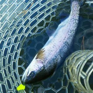
これが大正解。1投目からフェザージグにレインボーが、2投目クランクにも。
さらに1.6gスプーンにも釣れてきて、どうも中層で浮いている感じ。
お姉チャンもテンション上がって投げるも、木に引っかかったりで、なかなか掛からず。
しかし、ホスト役に徹してコチラが巻いて止めてを教えると早々にヒット!
その後も掛かる→ばらすを何回か繰り返し、充分楽しんで時間となりました。
結果、お姉チャンも釣れて自分も満足した釣果に良い釣りができました。
「良かったなぁ釣れて。また付いて行ってあげるわ」
と帰りの車で、お褒めの言葉を頂きました。


